Get trending papers in your email inbox once a day!
Get trending papers in your email inbox!
Subscribe
Conformal Risk Control for Pulmonary Nodule Detection
Quantitative tools are increasingly appealing for decision support in healthcare, driven by the growing capabilities of advanced AI systems. However, understanding the predictive uncertainties surrounding a tool's output is crucial for decision-makers to ensure reliable and transparent decisions. In this paper, we present a case study on pulmonary nodule detection for lung cancer screening, enhancing an advanced detection model with an uncertainty quantification technique called conformal risk control (CRC). We demonstrate that prediction sets with conformal guarantees are attractive measures of predictive uncertainty in the safety-critical healthcare domain, allowing end-users to achieve arbitrary validity by trading off false positives and providing formal statistical guarantees on model performance. Among ground-truth nodules annotated by at least three radiologists, our model achieves a sensitivity that is competitive with that generally achieved by individual radiologists, with a slight increase in false positives. Furthermore, we illustrate the risks of using off-the-shelve prediction models when faced with ontological uncertainty, such as when radiologists disagree on what constitutes the ground truth on pulmonary nodules.

AI in Lung Health: Benchmarking Detection and Diagnostic Models Across Multiple CT Scan Datasets
Lung cancer remains the leading cause of cancer-related mortality worldwide, and early detection through low-dose computed tomography (LDCT) has shown significant promise in reducing death rates. With the growing integration of artificial intelligence (AI) into medical imaging, the development and evaluation of robust AI models require access to large, well-annotated datasets. In this study, we introduce the utility of Duke Lung Cancer Screening (DLCS) Dataset, the largest open-access LDCT dataset with over 2,000 scans and 3,000 expert-verified nodules. We benchmark deep learning models for both 3D nodule detection and lung cancer classification across internal and external datasets including LUNA16, LUNA25, and NLST-3D+. For detection, we develop two MONAI-based RetinaNet models (DLCSDmD and LUNA16-mD), evaluated using the Competition Performance Metric (CPM). For classification, we compare five models, including state-of-the-art pretrained models (Models Genesis, Med3D), a selfsupervised foundation model (FMCB), a randomly initialized ResNet50, and proposed a novel Strategic Warm-Start++ (SWS++) model. SWS++ uses curated candidate patches to pretrain a classification backbone within the same detection pipeline, enabling task-relevant feature learning. Our models demonstrated strong generalizability, with SWS++ achieving comparable or superior performance to existing foundational models across multiple datasets (AUC: 0.71 to 0.90). All code, models, and data are publicly released to promote reproducibility and collaboration. This work establishes a standardized benchmarking resource for lung cancer AI research, supporting future efforts in model development, validation, and clinical translation.
T-SYNTH: A Knowledge-Based Dataset of Synthetic Breast Images
One of the key impediments for developing and assessing robust medical imaging algorithms is limited access to large-scale datasets with suitable annotations. Synthetic data generated with plausible physical and biological constraints may address some of these data limitations. We propose the use of physics simulations to generate synthetic images with pixel-level segmentation annotations, which are notoriously difficult to obtain. Specifically, we apply this approach to breast imaging analysis and release T-SYNTH, a large-scale open-source dataset of paired 2D digital mammography (DM) and 3D digital breast tomosynthesis (DBT) images. Our initial experimental results indicate that T-SYNTH images show promise for augmenting limited real patient datasets for detection tasks in DM and DBT. Our data and code are publicly available at https://github.com/DIDSR/tsynth-release.
Neural Graphics Primitives-based Deformable Image Registration for On-the-fly Motion Extraction
Intra-fraction motion in radiotherapy is commonly modeled using deformable image registration (DIR). However, existing methods often struggle to balance speed and accuracy, limiting their applicability in clinical scenarios. This study introduces a novel approach that harnesses Neural Graphics Primitives (NGP) to optimize the displacement vector field (DVF). Our method leverages learned primitives, processed as splats, and interpolates within space using a shallow neural network. Uniquely, it enables self-supervised optimization at an ultra-fast speed, negating the need for pre-training on extensive datasets and allowing seamless adaptation to new cases. We validated this approach on the 4D-CT lung dataset DIR-lab, achieving a target registration error (TRE) of 1.15\pm1.15 mm within a remarkable time of 1.77 seconds. Notably, our method also addresses the sliding boundary problem, a common challenge in conventional DIR methods.
SurGrID: Controllable Surgical Simulation via Scene Graph to Image Diffusion
Surgical simulation offers a promising addition to conventional surgical training. However, available simulation tools lack photorealism and rely on hardcoded behaviour. Denoising Diffusion Models are a promising alternative for high-fidelity image synthesis, but existing state-of-the-art conditioning methods fall short in providing precise control or interactivity over the generated scenes. We introduce SurGrID, a Scene Graph to Image Diffusion Model, allowing for controllable surgical scene synthesis by leveraging Scene Graphs. These graphs encode a surgical scene's components' spatial and semantic information, which are then translated into an intermediate representation using our novel pre-training step that explicitly captures local and global information. Our proposed method improves the fidelity of generated images and their coherence with the graph input over the state-of-the-art. Further, we demonstrate the simulation's realism and controllability in a user assessment study involving clinical experts. Scene Graphs can be effectively used for precise and interactive conditioning of Denoising Diffusion Models for simulating surgical scenes, enabling high fidelity and interactive control over the generated content.
A Lung Nodule Dataset with Histopathology-based Cancer Type Annotation
Recently, Computer-Aided Diagnosis (CAD) systems have emerged as indispensable tools in clinical diagnostic workflows, significantly alleviating the burden on radiologists. Nevertheless, despite their integration into clinical settings, CAD systems encounter limitations. Specifically, while CAD systems can achieve high performance in the detection of lung nodules, they face challenges in accurately predicting multiple cancer types. This limitation can be attributed to the scarcity of publicly available datasets annotated with expert-level cancer type information. This research aims to bridge this gap by providing publicly accessible datasets and reliable tools for medical diagnosis, facilitating a finer categorization of different types of lung diseases so as to offer precise treatment recommendations. To achieve this objective, we curated a diverse dataset of lung Computed Tomography (CT) images, comprising 330 annotated nodules (nodules are labeled as bounding boxes) from 95 distinct patients. The quality of the dataset was evaluated using a variety of classical classification and detection models, and these promising results demonstrate that the dataset has a feasible application and further facilitate intelligent auxiliary diagnosis.
SG2VID: Scene Graphs Enable Fine-Grained Control for Video Synthesis
Surgical simulation plays a pivotal role in training novice surgeons, accelerating their learning curve and reducing intra-operative errors. However, conventional simulation tools fall short in providing the necessary photorealism and the variability of human anatomy. In response, current methods are shifting towards generative model-based simulators. Yet, these approaches primarily focus on using increasingly complex conditioning for precise synthesis while neglecting the fine-grained human control aspect. To address this gap, we introduce SG2VID, the first diffusion-based video model that leverages Scene Graphs for both precise video synthesis and fine-grained human control. We demonstrate SG2VID's capabilities across three public datasets featuring cataract and cholecystectomy surgery. While SG2VID outperforms previous methods both qualitatively and quantitatively, it also enables precise synthesis, providing accurate control over tool and anatomy's size and movement, entrance of new tools, as well as the overall scene layout. We qualitatively motivate how SG2VID can be used for generative augmentation and present an experiment demonstrating its ability to improve a downstream phase detection task when the training set is extended with our synthetic videos. Finally, to showcase SG2VID's ability to retain human control, we interact with the Scene Graphs to generate new video samples depicting major yet rare intra-operative irregularities.
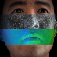
Relationship between pulmonary nodule malignancy and surrounding pleurae, airways and vessels: a quantitative study using the public LIDC-IDRI dataset
To investigate whether the pleurae, airways and vessels surrounding a nodule on non-contrast computed tomography (CT) can discriminate benign and malignant pulmonary nodules. The LIDC-IDRI dataset, one of the largest publicly available CT database, was exploited for study. A total of 1556 nodules from 694 patients were involved in statistical analysis, where nodules with average scorings <3 and >3 were respectively denoted as benign and malignant. Besides, 339 nodules from 113 patients with diagnosis ground-truth were independently evaluated. Computer algorithms were developed to segment pulmonary structures and quantify the distances to pleural surface, airways and vessels, as well as the counting number and normalized volume of airways and vessels near a nodule. Odds ratio (OR) and Chi-square (\chi^2) testing were performed to demonstrate the correlation between features of surrounding structures and nodule malignancy. A non-parametric receiver operating characteristic (ROC) analysis was conducted in logistic regression to evaluate discrimination ability of each structure. For benign and malignant groups, the average distances from nodules to pleural surface, airways and vessels are respectively (6.56, 5.19), (37.08, 26.43) and (1.42, 1.07) mm. The correlation between nodules and the counting number of airways and vessels that contact or project towards nodules are respectively (OR=22.96, \chi^2=105.04) and (OR=7.06, \chi^2=290.11). The correlation between nodules and the volume of airways and vessels are (OR=9.19, \chi^2=159.02) and (OR=2.29, \chi^2=55.89). The areas-under-curves (AUCs) for pleurae, airways and vessels are respectively 0.5202, 0.6943 and 0.6529. Our results show that malignant nodules are often surrounded by more pulmonary structures compared with benign ones, suggesting that features of these structures could be viewed as lung cancer biomarkers.

3DFPN-HS^2: 3D Feature Pyramid Network Based High Sensitivity and Specificity Pulmonary Nodule Detection
Accurate detection of pulmonary nodules with high sensitivity and specificity is essential for automatic lung cancer diagnosis from CT scans. Although many deep learning-based algorithms make great progress for improving the accuracy of nodule detection, the high false positive rate is still a challenging problem which limited the automatic diagnosis in routine clinical practice. In this paper, we propose a novel pulmonary nodule detection framework based on a 3D Feature Pyramid Network (3DFPN) to improve the sensitivity of nodule detection by employing multi-scale features to increase the resolution of nodules, as well as a parallel top-down path to transit the high-level semantic features to complement low-level general features. Furthermore, a High Sensitivity and Specificity (HS^2) network is introduced to eliminate the falsely detected nodule candidates by tracking the appearance changes in continuous CT slices of each nodule candidate. The proposed framework is evaluated on the public Lung Nodule Analysis (LUNA16) challenge dataset. Our method is able to accurately detect lung nodules at high sensitivity and specificity and achieves 90.4% sensitivity with 1/8 false positive per scan which outperforms the state-of-the-art results 15.6%.
Segmentation of 3D pore space from CT images using curvilinear skeleton: application to numerical simulation of microbial decomposition
Recent advances in 3D X-ray Computed Tomographic (CT) sensors have stimulated research efforts to unveil the extremely complex micro-scale processes that control the activity of soil microorganisms. Voxel-based description (up to hundreds millions voxels) of the pore space can be extracted, from grey level 3D CT scanner images, by means of simple image processing tools. Classical methods for numerical simulation of biological dynamics using mesh of voxels, such as Lattice Boltzmann Model (LBM), are too much time consuming. Thus, the use of more compact and reliable geometrical representations of pore space can drastically decrease the computational cost of the simulations. Several recent works propose basic analytic volume primitives (e.g. spheres, generalized cylinders, ellipsoids) to define a piece-wise approximation of pore space for numerical simulation of draining, diffusion and microbial decomposition. Such approaches work well but the drawback is that it generates approximation errors. In the present work, we study another alternative where pore space is described by means of geometrically relevant connected subsets of voxels (regions) computed from the curvilinear skeleton. Indeed, many works use the curvilinear skeleton (3D medial axis) for analyzing and partitioning 3D shapes within various domains (medicine, material sciences, petroleum engineering, etc.) but only a few ones in soil sciences. Within the context of soil sciences, most studies dealing with 3D medial axis focus on the determination of pore throats. Here, we segment pore space using curvilinear skeleton in order to achieve numerical simulation of microbial decomposition (including diffusion processes). We validate simulation outputs by comparison with other methods using different pore space geometrical representations (balls, voxels).
DDGS-CT: Direction-Disentangled Gaussian Splatting for Realistic Volume Rendering
Digitally reconstructed radiographs (DRRs) are simulated 2D X-ray images generated from 3D CT volumes, widely used in preoperative settings but limited in intraoperative applications due to computational bottlenecks, especially for accurate but heavy physics-based Monte Carlo methods. While analytical DRR renderers offer greater efficiency, they overlook anisotropic X-ray image formation phenomena, such as Compton scattering. We present a novel approach that marries realistic physics-inspired X-ray simulation with efficient, differentiable DRR generation using 3D Gaussian splatting (3DGS). Our direction-disentangled 3DGS (DDGS) method separates the radiosity contribution into isotropic and direction-dependent components, approximating complex anisotropic interactions without intricate runtime simulations. Additionally, we adapt the 3DGS initialization to account for tomography data properties, enhancing accuracy and efficiency. Our method outperforms state-of-the-art techniques in image accuracy. Furthermore, our DDGS shows promise for intraoperative applications and inverse problems such as pose registration, delivering superior registration accuracy and runtime performance compared to analytical DRR methods.

ROOM: A Physics-Based Continuum Robot Simulator for Photorealistic Medical Datasets Generation
Continuum robots are advancing bronchoscopy procedures by accessing complex lung airways and enabling targeted interventions. However, their development is limited by the lack of realistic training and test environments: Real data is difficult to collect due to ethical constraints and patient safety concerns, and developing autonomy algorithms requires realistic imaging and physical feedback. We present ROOM (Realistic Optical Observation in Medicine), a comprehensive simulation framework designed for generating photorealistic bronchoscopy training data. By leveraging patient CT scans, our pipeline renders multi-modal sensor data including RGB images with realistic noise and light specularities, metric depth maps, surface normals, optical flow and point clouds at medically relevant scales. We validate the data generated by ROOM in two canonical tasks for medical robotics -- multi-view pose estimation and monocular depth estimation, demonstrating diverse challenges that state-of-the-art methods must overcome to transfer to these medical settings. Furthermore, we show that the data produced by ROOM can be used to fine-tune existing depth estimation models to overcome these challenges, also enabling other downstream applications such as navigation. We expect that ROOM will enable large-scale data generation across diverse patient anatomies and procedural scenarios that are challenging to capture in clinical settings. Code and data: https://github.com/iamsalvatore/room.

How to Trust Your Diffusion Model: A Convex Optimization Approach to Conformal Risk Control
Score-based generative modeling, informally referred to as diffusion models, continue to grow in popularity across several important domains and tasks. While they provide high-quality and diverse samples from empirical distributions, important questions remain on the reliability and trustworthiness of these sampling procedures for their responsible use in critical scenarios. Conformal prediction is a modern tool to construct finite-sample, distribution-free uncertainty guarantees for any black-box predictor. In this work, we focus on image-to-image regression tasks and we present a generalization of the Risk-Controlling Prediction Sets (RCPS) procedure, that we term K-RCPS, which allows to (i) provide entrywise calibrated intervals for future samples of any diffusion model, and (ii) control a certain notion of risk with respect to a ground truth image with minimal mean interval length. Differently from existing conformal risk control procedures, ours relies on a novel convex optimization approach that allows for multidimensional risk control while provably minimizing the mean interval length. We illustrate our approach on two real-world image denoising problems: on natural images of faces as well as on computed tomography (CT) scans of the abdomen, demonstrating state of the art performance.

LeFusion: Controllable Pathology Synthesis via Lesion-Focused Diffusion Models
Patient data from real-world clinical practice often suffers from data scarcity and long-tail imbalances, leading to biased outcomes or algorithmic unfairness. This study addresses these challenges by generating lesion-containing image-segmentation pairs from lesion-free images. Previous efforts in medical imaging synthesis have struggled with separating lesion information from background, resulting in low-quality backgrounds and limited control over the synthetic output. Inspired by diffusion-based image inpainting, we propose LeFusion, a lesion-focused diffusion model. By redesigning the diffusion learning objectives to focus on lesion areas, we simplify the learning process and improve control over the output while preserving high-fidelity backgrounds by integrating forward-diffused background contexts into the reverse diffusion process. Additionally, we tackle two major challenges in lesion texture synthesis: 1) multi-peak and 2) multi-class lesions. We introduce two effective strategies: histogram-based texture control and multi-channel decomposition, enabling the controlled generation of high-quality lesions in difficult scenarios. Furthermore, we incorporate lesion mask diffusion, allowing control over lesion size, location, and boundary, thus increasing lesion diversity. Validated on 3D cardiac lesion MRI and lung nodule CT datasets, LeFusion-generated data significantly improves the performance of state-of-the-art segmentation models, including nnUNet and SwinUNETR. Code and model are available at https://github.com/M3DV/LeFusion.


Suturing Tasks Automation Based on Skills Learned From Demonstrations: A Simulation Study
In this work, we develop an open-source surgical simulation environment that includes a realistic model obtained by MRI-scanning a physical phantom, for the purpose of training and evaluating a Learning from Demonstration (LfD) algorithm for autonomous suturing. The LfD algorithm utilizes Dynamic Movement Primitives (DMP) and Locally Weighted Regression (LWR), but focuses on the needle trajectory, rather than the instruments, to obtain better generality with respect to needle grasps. We conduct a user study to collect multiple suturing demonstrations and perform a comprehensive analysis of the ability of the LfD algorithm to generalize from a demonstration at one location in one phantom to different locations in the same phantom and to a different phantom. Our results indicate good generalization, on the order of 91.5%, when learning from more experienced subjects, indicating the need to integrate skill assessment in the future.
Peritumoral Expansion Radiomics for Improved Lung Cancer Classification
Purpose: This study investigated how nodule segmentation and surrounding peritumoral regions influence radionics-based lung cancer classification. Methods: Using 3D CT scans with bounding box annotated nodules, we generated 3D segmentations using four techniques: Otsu, Fuzzy C-Means (FCM), Gaussian Mixture Model (GMM), and K-Nearest Neighbors (KNN). Radiomics features were extracted using the PyRadiomics library, and multiple machine-learning-based classifiers, including Random Forest, Logistic Regression, and KNN, were employed to classify nodules as cancerous or non-cancerous. The best-performing segmentation and model were further analyzed by expanding the initial nodule segmentation into the peritumoral region (2, 4, 6, 8, 10, and 12 mm) to understand the influence of the surrounding area on classification. Additionally, we compared our results to deep learning-based feature extractors Foundation Model for Cancer Biomarkers (FMCB) and other state-of-the-art baseline models. Results: Incorporating peritumoral regions significantly enhanced performance, with the best result obtained at 8 mm expansion (AUC = 0.78). Compared to image-based deep learning models, such as FMCB (AUC = 0.71) and ResNet50-SWS++ (AUC = 0.71), our radiomics-based approach demonstrated superior classification accuracy. Conclusion: The study highlights the importance of peritumoral expansion in improving lung cancer classification using radiomics. These findings can inform the development of more robust AI-driven diagnostic tools.
Knowledge-based in silico models and dataset for the comparative evaluation of mammography AI for a range of breast characteristics, lesion conspicuities and doses
To generate evidence regarding the safety and efficacy of artificial intelligence (AI) enabled medical devices, AI models need to be evaluated on a diverse population of patient cases, some of which may not be readily available. We propose an evaluation approach for testing medical imaging AI models that relies on in silico imaging pipelines in which stochastic digital models of human anatomy (in object space) with and without pathology are imaged using a digital replica imaging acquisition system to generate realistic synthetic image datasets. Here, we release M-SYNTH, a dataset of cohorts with four breast fibroglandular density distributions imaged at different exposure levels using Monte Carlo x-ray simulations with the publicly available Virtual Imaging Clinical Trial for Regulatory Evaluation (VICTRE) toolkit. We utilize the synthetic dataset to analyze AI model performance and find that model performance decreases with increasing breast density and increases with higher mass density, as expected. As exposure levels decrease, AI model performance drops with the highest performance achieved at exposure levels lower than the nominal recommended dose for the breast type.
Head and Neck Tumor Segmentation from [18F]F-FDG PET/CT Images Based on 3D Diffusion Model
Head and neck (H&N) cancers are among the most prevalent types of cancer worldwide, and [18F]F-FDG PET/CT is widely used for H&N cancer management. Recently, the diffusion model has demonstrated remarkable performance in various image-generation tasks. In this work, we proposed a 3D diffusion model to accurately perform H&N tumor segmentation from 3D PET and CT volumes. The 3D diffusion model was developed considering the 3D nature of PET and CT images acquired. During the reverse process, the model utilized a 3D UNet structure and took the concatenation of PET, CT, and Gaussian noise volumes as the network input to generate the tumor mask. Experiments based on the HECKTOR challenge dataset were conducted to evaluate the effectiveness of the proposed diffusion model. Several state-of-the-art techniques based on U-Net and Transformer structures were adopted as the reference methods. Benefits of employing both PET and CT as the network input as well as further extending the diffusion model from 2D to 3D were investigated based on various quantitative metrics and the uncertainty maps generated. Results showed that the proposed 3D diffusion model could generate more accurate segmentation results compared with other methods. Compared to the diffusion model in 2D format, the proposed 3D model yielded superior results. Our experiments also highlighted the advantage of utilizing dual-modality PET and CT data over only single-modality data for H&N tumor segmentation.
MedSyn: Text-guided Anatomy-aware Synthesis of High-Fidelity 3D CT Images
This paper introduces an innovative methodology for producing high-quality 3D lung CT images guided by textual information. While diffusion-based generative models are increasingly used in medical imaging, current state-of-the-art approaches are limited to low-resolution outputs and underutilize radiology reports' abundant information. The radiology reports can enhance the generation process by providing additional guidance and offering fine-grained control over the synthesis of images. Nevertheless, expanding text-guided generation to high-resolution 3D images poses significant memory and anatomical detail-preserving challenges. Addressing the memory issue, we introduce a hierarchical scheme that uses a modified UNet architecture. We start by synthesizing low-resolution images conditioned on the text, serving as a foundation for subsequent generators for complete volumetric data. To ensure the anatomical plausibility of the generated samples, we provide further guidance by generating vascular, airway, and lobular segmentation masks in conjunction with the CT images. The model demonstrates the capability to use textual input and segmentation tasks to generate synthesized images. The results of comparative assessments indicate that our approach exhibits superior performance compared to the most advanced models based on GAN and diffusion techniques, especially in accurately retaining crucial anatomical features such as fissure lines, airways, and vascular structures. This innovation introduces novel possibilities. This study focuses on two main objectives: (1) the development of a method for creating images based on textual prompts and anatomical components, and (2) the capability to generate new images conditioning on anatomical elements. The advancements in image generation can be applied to enhance numerous downstream tasks.


Standardized Benchmark Dataset for Localized Exposure to a Realistic Source at 10-90 GHz
The lack of freely available standardized datasets represents an aggravating factor during the development and testing the performance of novel computational techniques in exposure assessment and dosimetry research. This hinders progress as researchers are required to generate numerical data (field, power and temperature distribution) anew using simulation software for each exposure scenario. Other than being time consuming, this approach is highly susceptible to errors that occur during the configuration of the electromagnetic model. To address this issue, in this paper, the limited available data on the incident power density and resultant maximum temperature rise on the skin surface considering various steady-state exposure scenarios at 10-90 GHz have been statistically modeled. The synthetic data have been sampled from the fitted statistical multivariate distribution with respect to predetermined dosimetric constraints. We thus present a comprehensive and open-source dataset compiled of the high-fidelity numerical data considering various exposures to a realistic source. Furthermore, different surrogate models for predicting maximum temperature rise on the skin surface were fitted based on the synthetic dataset. All surrogate models were tested on the originally available data where satisfactory predictive performance has been demonstrated. A simple technique of combining quadratic polynomial and tensor-product spline surrogates, each operating on its own cluster of data, has achieved the lowest mean absolute error of 0.058 {\deg}C. Therefore, overall experimental results indicate the validity of the proposed synthetic dataset.
OCTCube-M: A 3D multimodal optical coherence tomography foundation model for retinal and systemic diseases with cross-cohort and cross-device validation
We present OCTCube-M, a 3D OCT-based multi-modal foundation model for jointly analyzing OCT and en face images. OCTCube-M first developed OCTCube, a 3D foundation model pre-trained on 26,685 3D OCT volumes encompassing 1.62 million 2D OCT images. It then exploits a novel multi-modal contrastive learning framework COEP to integrate other retinal imaging modalities, such as fundus autofluorescence and infrared retinal imaging, into OCTCube, efficiently extending it into multi-modal foundation models. OCTCube achieves best performance on predicting 8 retinal diseases, demonstrating strong generalizability on cross-cohort, cross-device and cross-modality prediction. OCTCube can also predict cross-organ nodule malignancy (CT) and low cardiac ejection fraction as well as systemic diseases, such as diabetes and hypertension, revealing its wide applicability beyond retinal diseases. We further develop OCTCube-IR using COEP with 26,685 OCT and IR image pairs. OCTCube-IR can accurately retrieve between OCT and IR images, allowing joint analysis between 3D and 2D retinal imaging modalities. Finally, we trained a tri-modal foundation model OCTCube-EF from 4 million 2D OCT images and 400K en face retinal images. OCTCube-EF attains the best performance on predicting the growth rate of geographic atrophy (GA) across datasets collected from 6 multi-center global trials conducted in 23 countries. This improvement is statistically equivalent to running a clinical trial with more than double the size of the original study. Our analysis based on another retrospective case study reveals OCTCube-EF's ability to avoid false positive Phase-III results according to its accurate treatment effect estimation on the Phase-II results. In sum, OCTCube-M is a 3D multi-modal foundation model framework that integrates OCT and other retinal imaging modalities revealing substantial diagnostic and prognostic benefits.
AnyStar: Domain randomized universal star-convex 3D instance segmentation
Star-convex shapes arise across bio-microscopy and radiology in the form of nuclei, nodules, metastases, and other units. Existing instance segmentation networks for such structures train on densely labeled instances for each dataset, which requires substantial and often impractical manual annotation effort. Further, significant reengineering or finetuning is needed when presented with new datasets and imaging modalities due to changes in contrast, shape, orientation, resolution, and density. We present AnyStar, a domain-randomized generative model that simulates synthetic training data of blob-like objects with randomized appearance, environments, and imaging physics to train general-purpose star-convex instance segmentation networks. As a result, networks trained using our generative model do not require annotated images from unseen datasets. A single network trained on our synthesized data accurately 3D segments C. elegans and P. dumerilii nuclei in fluorescence microscopy, mouse cortical nuclei in micro-CT, zebrafish brain nuclei in EM, and placental cotyledons in human fetal MRI, all without any retraining, finetuning, transfer learning, or domain adaptation. Code is available at https://github.com/neel-dey/AnyStar.

Multi-Task Lung Nodule Detection in Chest Radiographs with a Dual Head Network
Lung nodules can be an alarming precursor to potential lung cancer. Missed nodule detections during chest radiograph analysis remains a common challenge among thoracic radiologists. In this work, we present a multi-task lung nodule detection algorithm for chest radiograph analysis. Unlike past approaches, our algorithm predicts a global-level label indicating nodule presence along with local-level labels predicting nodule locations using a Dual Head Network (DHN). We demonstrate the favorable nodule detection performance that our multi-task formulation yields in comparison to conventional methods. In addition, we introduce a novel Dual Head Augmentation (DHA) strategy tailored for DHN, and we demonstrate its significance in further enhancing global and local nodule predictions.
ZeroAvatar: Zero-shot 3D Avatar Generation from a Single Image
Recent advancements in text-to-image generation have enabled significant progress in zero-shot 3D shape generation. This is achieved by score distillation, a methodology that uses pre-trained text-to-image diffusion models to optimize the parameters of a 3D neural presentation, e.g. Neural Radiance Field (NeRF). While showing promising results, existing methods are often not able to preserve the geometry of complex shapes, such as human bodies. To address this challenge, we present ZeroAvatar, a method that introduces the explicit 3D human body prior to the optimization process. Specifically, we first estimate and refine the parameters of a parametric human body from a single image. Then during optimization, we use the posed parametric body as additional geometry constraint to regularize the diffusion model as well as the underlying density field. Lastly, we propose a UV-guided texture regularization term to further guide the completion of texture on invisible body parts. We show that ZeroAvatar significantly enhances the robustness and 3D consistency of optimization-based image-to-3D avatar generation, outperforming existing zero-shot image-to-3D methods.

Towards a clinically accessible radiology foundation model: open-access and lightweight, with automated evaluation
The scaling laws and extraordinary performance of large foundation models motivate the development and utilization of such models in biomedicine. However, despite early promising results on some biomedical benchmarks, there are still major challenges that need to be addressed before these models can be used in real-world clinics. Frontier general-domain models such as GPT-4V still have significant performance gaps in multimodal biomedical applications. More importantly, less-acknowledged pragmatic issues, including accessibility, model cost, and tedious manual evaluation make it hard for clinicians to use state-of-the-art large models directly on private patient data. Here, we explore training open-source small multimodal models (SMMs) to bridge competency gaps for unmet clinical needs in radiology. To maximize data efficiency, we adopt a modular approach by incorporating state-of-the-art pre-trained models for image and text modalities, and focusing on training a lightweight adapter to ground each modality to the text embedding space, as exemplified by LLaVA-Med. For training, we assemble a large dataset of over 697 thousand radiology image-text pairs. For evaluation, we propose CheXprompt, a GPT-4-based metric for factuality evaluation, and demonstrate its parity with expert evaluation. For best practice, we conduct a systematic ablation study on various choices in data engineering and multimodal training. The resulting LlaVA-Rad (7B) model attains state-of-the-art results on standard radiology tasks such as report generation and cross-modal retrieval, even outperforming much larger models such as GPT-4V and Med-PaLM M (84B). The inference of LlaVA-Rad is fast and can be performed on a single V100 GPU in private settings, offering a promising state-of-the-art tool for real-world clinical applications.


Non-Invasive Medical Digital Twins using Physics-Informed Self-Supervised Learning
A digital twin is a virtual replica of a real-world physical phenomena that uses mathematical modeling to characterize and simulate its defining features. By constructing digital twins for disease processes, we can perform in-silico simulations that mimic patients' health conditions and counterfactual outcomes under hypothetical interventions in a virtual setting. This eliminates the need for invasive procedures or uncertain treatment decisions. In this paper, we propose a method to identify digital twin model parameters using only noninvasive patient health data. We approach the digital twin modeling as a composite inverse problem, and observe that its structure resembles pretraining and finetuning in self-supervised learning (SSL). Leveraging this, we introduce a physics-informed SSL algorithm that initially pretrains a neural network on the pretext task of solving the physical model equations. Subsequently, the model is trained to reconstruct low-dimensional health measurements from noninvasive modalities while being constrained by the physical equations learned in pretraining. We apply our method to identify digital twins of cardiac hemodynamics using noninvasive echocardiogram videos, and demonstrate its utility in unsupervised disease detection and in-silico clinical trials.

Medical World Model: Generative Simulation of Tumor Evolution for Treatment Planning
Providing effective treatment and making informed clinical decisions are essential goals of modern medicine and clinical care. We are interested in simulating disease dynamics for clinical decision-making, leveraging recent advances in large generative models. To this end, we introduce the Medical World Model (MeWM), the first world model in medicine that visually predicts future disease states based on clinical decisions. MeWM comprises (i) vision-language models to serve as policy models, and (ii) tumor generative models as dynamics models. The policy model generates action plans, such as clinical treatments, while the dynamics model simulates tumor progression or regression under given treatment conditions. Building on this, we propose the inverse dynamics model that applies survival analysis to the simulated post-treatment tumor, enabling the evaluation of treatment efficacy and the selection of the optimal clinical action plan. As a result, the proposed MeWM simulates disease dynamics by synthesizing post-treatment tumors, with state-of-the-art specificity in Turing tests evaluated by radiologists. Simultaneously, its inverse dynamics model outperforms medical-specialized GPTs in optimizing individualized treatment protocols across all metrics. Notably, MeWM improves clinical decision-making for interventional physicians, boosting F1-score in selecting the optimal TACE protocol by 13%, paving the way for future integration of medical world models as the second readers.
A Cross Spatio-Temporal Pathology-based Lung Nodule Dataset
Recently, intelligent analysis of lung nodules with the assistant of computer aided detection (CAD) techniques can improve the accuracy rate of lung cancer diagnosis. However, existing CAD systems and pulmonary datasets mainly focus on Computed Tomography (CT) images from one single period, while ignoring the cross spatio-temporal features associated with the progression of nodules contained in imaging data from various captured periods of lung cancer. If the evolution patterns of nodules across various periods in the patients' CT sequences can be explored, it will play a crucial role in guiding the precise screening identification of lung cancer. Therefore, a cross spatio-temporal lung nodule dataset with pathological information for nodule identification and diagnosis is constructed, which contains 328 CT sequences and 362 annotated nodules from 109 patients. This comprehensive database is intended to drive research in the field of CAD towards more practical and robust methods, and also contribute to the further exploration of precision medicine related field. To ensure patient confidentiality, we have removed sensitive information from the dataset.
Text-Driven Tumor Synthesis
Tumor synthesis can generate examples that AI often misses or over-detects, improving AI performance by training on these challenging cases. However, existing synthesis methods, which are typically unconditional -- generating images from random variables -- or conditioned only by tumor shapes, lack controllability over specific tumor characteristics such as texture, heterogeneity, boundaries, and pathology type. As a result, the generated tumors may be overly similar or duplicates of existing training data, failing to effectively address AI's weaknesses. We propose a new text-driven tumor synthesis approach, termed TextoMorph, that provides textual control over tumor characteristics. This is particularly beneficial for examples that confuse the AI the most, such as early tumor detection (increasing Sensitivity by +8.5%), tumor segmentation for precise radiotherapy (increasing DSC by +6.3%), and classification between benign and malignant tumors (improving Sensitivity by +8.2%). By incorporating text mined from radiology reports into the synthesis process, we increase the variability and controllability of the synthetic tumors to target AI's failure cases more precisely. Moreover, TextoMorph uses contrastive learning across different texts and CT scans, significantly reducing dependence on scarce image-report pairs (only 141 pairs used in this study) by leveraging a large corpus of 34,035 radiology reports. Finally, we have developed rigorous tests to evaluate synthetic tumors, including Text-Driven Visual Turing Test and Radiomics Pattern Analysis, showing that our synthetic tumors is realistic and diverse in texture, heterogeneity, boundaries, and pathology.


PIE: Simulating Disease Progression via Progressive Image Editing
Disease progression simulation is a crucial area of research that has significant implications for clinical diagnosis, prognosis, and treatment. One major challenge in this field is the lack of continuous medical imaging monitoring of individual patients over time. To address this issue, we develop a novel framework termed Progressive Image Editing (PIE) that enables controlled manipulation of disease-related image features, facilitating precise and realistic disease progression simulation. Specifically, we leverage recent advancements in text-to-image generative models to simulate disease progression accurately and personalize it for each patient. We theoretically analyze the iterative refining process in our framework as a gradient descent with an exponentially decayed learning rate. To validate our framework, we conduct experiments in three medical imaging domains. Our results demonstrate the superiority of PIE over existing methods such as Stable Diffusion Walk and Style-Based Manifold Extrapolation based on CLIP score (Realism) and Disease Classification Confidence (Alignment). Our user study collected feedback from 35 veteran physicians to assess the generated progressions. Remarkably, 76.2% of the feedback agrees with the fidelity of the generated progressions. To our best knowledge, PIE is the first of its kind to generate disease progression images meeting real-world standards. It is a promising tool for medical research and clinical practice, potentially allowing healthcare providers to model disease trajectories over time, predict future treatment responses, and improve patient outcomes.


Bora: Biomedical Generalist Video Generation Model
Generative models hold promise for revolutionizing medical education, robot-assisted surgery, and data augmentation for medical AI development. Diffusion models can now generate realistic images from text prompts, while recent advancements have demonstrated their ability to create diverse, high-quality videos. However, these models often struggle with generating accurate representations of medical procedures and detailed anatomical structures. This paper introduces Bora, the first spatio-temporal diffusion probabilistic model designed for text-guided biomedical video generation. Bora leverages Transformer architecture and is pre-trained on general-purpose video generation tasks. It is fine-tuned through model alignment and instruction tuning using a newly established medical video corpus, which includes paired text-video data from various biomedical fields. To the best of our knowledge, this is the first attempt to establish such a comprehensive annotated biomedical video dataset. Bora is capable of generating high-quality video data across four distinct biomedical domains, adhering to medical expert standards and demonstrating consistency and diversity. This generalist video generative model holds significant potential for enhancing medical consultation and decision-making, particularly in resource-limited settings. Additionally, Bora could pave the way for immersive medical training and procedure planning. Extensive experiments on distinct medical modalities such as endoscopy, ultrasound, MRI, and cell tracking validate the effectiveness of our model in understanding biomedical instructions and its superior performance across subjects compared to state-of-the-art generation models.

Real-Time Neural Rasterization for Large Scenes
We propose a new method for realistic real-time novel-view synthesis (NVS) of large scenes. Existing neural rendering methods generate realistic results, but primarily work for small scale scenes (<50 square meters) and have difficulty at large scale (>10000 square meters). Traditional graphics-based rasterization rendering is fast for large scenes but lacks realism and requires expensive manually created assets. Our approach combines the best of both worlds by taking a moderate-quality scaffold mesh as input and learning a neural texture field and shader to model view-dependant effects to enhance realism, while still using the standard graphics pipeline for real-time rendering. Our method outperforms existing neural rendering methods, providing at least 30x faster rendering with comparable or better realism for large self-driving and drone scenes. Our work is the first to enable real-time rendering of large real-world scenes.
S-SYNTH: Knowledge-Based, Synthetic Generation of Skin Images
Development of artificial intelligence (AI) techniques in medical imaging requires access to large-scale and diverse datasets for training and evaluation. In dermatology, obtaining such datasets remains challenging due to significant variations in patient populations, illumination conditions, and acquisition system characteristics. In this work, we propose S-SYNTH, the first knowledge-based, adaptable open-source skin simulation framework to rapidly generate synthetic skin, 3D models and digitally rendered images, using an anatomically inspired multi-layer, multi-component skin and growing lesion model. The skin model allows for controlled variation in skin appearance, such as skin color, presence of hair, lesion shape, and blood fraction among other parameters. We use this framework to study the effect of possible variations on the development and evaluation of AI models for skin lesion segmentation, and show that results obtained using synthetic data follow similar comparative trends as real dermatologic images, while mitigating biases and limitations from existing datasets including small dataset size, lack of diversity, and underrepresentation.
EndoPBR: Material and Lighting Estimation for Photorealistic Surgical Simulations via Physically-based Rendering
The lack of labeled datasets in 3D vision for surgical scenes inhibits the development of robust 3D reconstruction algorithms in the medical domain. Despite the popularity of Neural Radiance Fields and 3D Gaussian Splatting in the general computer vision community, these systems have yet to find consistent success in surgical scenes due to challenges such as non-stationary lighting and non-Lambertian surfaces. As a result, the need for labeled surgical datasets continues to grow. In this work, we introduce a differentiable rendering framework for material and lighting estimation from endoscopic images and known geometry. Compared to previous approaches that model lighting and material jointly as radiance, we explicitly disentangle these scene properties for robust and photorealistic novel view synthesis. To disambiguate the training process, we formulate domain-specific properties inherent in surgical scenes. Specifically, we model the scene lighting as a simple spotlight and material properties as a bidirectional reflectance distribution function, parameterized by a neural network. By grounding color predictions in the rendering equation, we can generate photorealistic images at arbitrary camera poses. We evaluate our method with various sequences from the Colonoscopy 3D Video Dataset and show that our method produces competitive novel view synthesis results compared with other approaches. Furthermore, we demonstrate that synthetic data can be used to develop 3D vision algorithms by finetuning a depth estimation model with our rendered outputs. Overall, we see that the depth estimation performance is on par with fine-tuning with the original real images.
EndoGaussian: Real-time Gaussian Splatting for Dynamic Endoscopic Scene Reconstruction
Reconstructing deformable tissues from endoscopic videos is essential in many downstream surgical applications. However, existing methods suffer from slow rendering speed, greatly limiting their practical use. In this paper, we introduce EndoGaussian, a real-time endoscopic scene reconstruction framework built on 3D Gaussian Splatting (3DGS). By integrating the efficient Gaussian representation and highly-optimized rendering engine, our framework significantly boosts the rendering speed to a real-time level. To adapt 3DGS for endoscopic scenes, we propose two strategies, Holistic Gaussian Initialization (HGI) and Spatio-temporal Gaussian Tracking (SGT), to handle the non-trivial Gaussian initialization and tissue deformation problems, respectively. In HGI, we leverage recent depth estimation models to predict depth maps of input binocular/monocular image sequences, based on which pixels are re-projected and combined for holistic initialization. In SPT, we propose to model surface dynamics using a deformation field, which is composed of an efficient encoding voxel and a lightweight deformation decoder, allowing for Gaussian tracking with minor training and rendering burden. Experiments on public datasets demonstrate our efficacy against prior SOTAs in many aspects, including better rendering speed (195 FPS real-time, 100times gain), better rendering quality (37.848 PSNR), and less training overhead (within 2 min/scene), showing significant promise for intraoperative surgery applications. Code is available at: https://yifliu3.github.io/EndoGaussian/.
A for-loop is all you need. For solving the inverse problem in the case of personalized tumor growth modeling
Solving the inverse problem is the key step in evaluating the capacity of a physical model to describe real phenomena. In medical image computing, it aligns with the classical theme of image-based model personalization. Traditionally, a solution to the problem is obtained by performing either sampling or variational inference based methods. Both approaches aim to identify a set of free physical model parameters that results in a simulation best matching an empirical observation. When applied to brain tumor modeling, one of the instances of image-based model personalization in medical image computing, the overarching drawback of the methods is the time complexity for finding such a set. In a clinical setting with limited time between imaging and diagnosis or even intervention, this time complexity may prove critical. As the history of quantitative science is the history of compression, we align in this paper with the historical tendency and propose a method compressing complex traditional strategies for solving an inverse problem into a simple database query task. We evaluated different ways of performing the database query task assessing the trade-off between accuracy and execution time. On the exemplary task of brain tumor growth modeling, we prove that the proposed method achieves one order speed-up compared to existing approaches for solving the inverse problem. The resulting compute time offers critical means for relying on more complex and, hence, realistic models, for integrating image preprocessing and inverse modeling even deeper, or for implementing the current model into a clinical workflow.
Leveraging Semantic Asymmetry for Precise Gross Tumor Volume Segmentation of Nasopharyngeal Carcinoma in Planning CT
In the radiation therapy of nasopharyngeal carcinoma (NPC), clinicians typically delineate the gross tumor volume (GTV) using non-contrast planning computed tomography to ensure accurate radiation dose delivery. However, the low contrast between tumors and adjacent normal tissues necessitates that radiation oncologists manually delineate the tumors, often relying on diagnostic MRI for guidance. % In this study, we propose a novel approach to directly segment NPC gross tumors on non-contrast planning CT images, circumventing potential registration errors when aligning MRI or MRI-derived tumor masks to planning CT. To address the low contrast issues between tumors and adjacent normal structures in planning CT, we introduce a 3D Semantic Asymmetry Tumor segmentation (SATs) method. Specifically, we posit that a healthy nasopharyngeal region is characteristically bilaterally symmetric, whereas the emergence of nasopharyngeal carcinoma disrupts this symmetry. Then, we propose a Siamese contrastive learning segmentation framework that minimizes the voxel-wise distance between original and flipped areas without tumor and encourages a larger distance between original and flipped areas with tumor. Thus, our approach enhances the sensitivity of features to semantic asymmetries. % Extensive experiments demonstrate that the proposed SATs achieves the leading NPC GTV segmentation performance in both internal and external testing, e.g., with at least 2\% absolute Dice score improvement and 12\% average distance error reduction when compared to other state-of-the-art methods in the external testing.
Towards Generalist Foundation Model for Radiology
In this study, we aim to initiate the development of Radiology Foundation Model, termed as RadFM.We consider the construction of foundational models from the perspectives of data, model design, and evaluation thoroughly. Our contribution can be concluded as follows: (i), we construct a large-scale Medical Multi-modal Dataset, MedMD, consisting of 16M 2D and 3D medical scans. To the best of our knowledge, this is the first multi-modal dataset containing 3D medical scans. (ii), We propose an architecture that enables visually conditioned generative pre-training, allowing for the integration of text input interleaved with 2D or 3D medical scans to generate response for diverse radiologic tasks. The model was initially pre-trained on MedMD and subsequently domain-specific fine-tuned on RadMD, a radiologic cleaned version of MedMD, containing 3M radiologic visual-language pairs. (iii), we propose a new evaluation benchmark that comprises five tasks, aiming to comprehensively assess the capability of foundation models in handling practical clinical problems. Our experimental results confirm that RadFM significantly outperforms existing multi-modal foundation models. The codes, data, and model checkpoint will all be made publicly available to promote further research and development in the field.

Adaptive Shells for Efficient Neural Radiance Field Rendering
Neural radiance fields achieve unprecedented quality for novel view synthesis, but their volumetric formulation remains expensive, requiring a huge number of samples to render high-resolution images. Volumetric encodings are essential to represent fuzzy geometry such as foliage and hair, and they are well-suited for stochastic optimization. Yet, many scenes ultimately consist largely of solid surfaces which can be accurately rendered by a single sample per pixel. Based on this insight, we propose a neural radiance formulation that smoothly transitions between volumetric- and surface-based rendering, greatly accelerating rendering speed and even improving visual fidelity. Our method constructs an explicit mesh envelope which spatially bounds a neural volumetric representation. In solid regions, the envelope nearly converges to a surface and can often be rendered with a single sample. To this end, we generalize the NeuS formulation with a learned spatially-varying kernel size which encodes the spread of the density, fitting a wide kernel to volume-like regions and a tight kernel to surface-like regions. We then extract an explicit mesh of a narrow band around the surface, with width determined by the kernel size, and fine-tune the radiance field within this band. At inference time, we cast rays against the mesh and evaluate the radiance field only within the enclosed region, greatly reducing the number of samples required. Experiments show that our approach enables efficient rendering at very high fidelity. We also demonstrate that the extracted envelope enables downstream applications such as animation and simulation.


Rapid patient-specific neural networks for intraoperative X-ray to volume registration
The integration of artificial intelligence in image-guided interventions holds transformative potential, promising to extract 3D geometric and quantitative information from conventional 2D imaging modalities during complex procedures. Achieving this requires the rapid and precise alignment of 2D intraoperative images (e.g., X-ray) with 3D preoperative volumes (e.g., CT, MRI). However, current 2D/3D registration methods fail across the broad spectrum of procedures dependent on X-ray guidance: traditional optimization techniques require custom parameter tuning for each subject, whereas neural networks trained on small datasets do not generalize to new patients or require labor-intensive manual annotations, increasing clinical burden and precluding application to new anatomical targets. To address these challenges, we present xvr, a fully automated framework for training patient-specific neural networks for 2D/3D registration. xvr uses physics-based simulation to generate abundant high-quality training data from a patient's own preoperative volumetric imaging, thereby overcoming the inherently limited ability of supervised models to generalize to new patients and procedures. Furthermore, xvr requires only 5 minutes of training per patient, making it suitable for emergency interventions as well as planned procedures. We perform the largest evaluation of a 2D/3D registration algorithm on real X-ray data to date and find that xvr robustly generalizes across a diverse dataset comprising multiple anatomical structures, imaging modalities, and hospitals. Across surgical tasks, xvr achieves submillimeter-accurate registration at intraoperative speeds, improving upon existing methods by an order of magnitude. xvr is released as open-source software freely available at https://github.com/eigenvivek/xvr.

Individualizing Glioma Radiotherapy Planning by Optimization of Data and Physics-Informed Discrete Loss
Brain tumor growth is unique to each glioma patient and extends beyond what is visible in imaging scans, infiltrating surrounding brain tissue. Understanding these hidden patient-specific progressions is essential for effective therapies. Current treatment plans for brain tumors, such as radiotherapy, typically involve delineating a uniform margin around the visible tumor on pre-treatment scans to target this invisible tumor growth. This "one size fits all" approach is derived from population studies and often fails to account for the nuances of individual patient conditions. We present the GliODIL framework, which infers the full spatial distribution of tumor cell concentration from available multi-modal imaging, leveraging a Fisher-Kolmogorov type physics model to describe tumor growth. This is achieved through the newly introduced method of Optimizing the Discrete Loss (ODIL), where both data and physics-based constraints are softly assimilated into the solution. Our test dataset comprises 152 glioblastoma patients with pre-treatment imaging and post-treatment follow-ups for tumor recurrence monitoring. By blending data-driven techniques with physics-based constraints, GliODIL enhances recurrence prediction in radiotherapy planning, challenging traditional uniform margins and strict adherence to the Fisher-Kolmogorov partial differential equation (PDE) model, which is adapted for complex cases.
Label-Free Liver Tumor Segmentation
We demonstrate that AI models can accurately segment liver tumors without the need for manual annotation by using synthetic tumors in CT scans. Our synthetic tumors have two intriguing advantages: (I) realistic in shape and texture, which even medical professionals can confuse with real tumors; (II) effective for training AI models, which can perform liver tumor segmentation similarly to the model trained on real tumors -- this result is exciting because no existing work, using synthetic tumors only, has thus far reached a similar or even close performance to real tumors. This result also implies that manual efforts for annotating tumors voxel by voxel (which took years to create) can be significantly reduced in the future. Moreover, our synthetic tumors can automatically generate many examples of small (or even tiny) synthetic tumors and have the potential to improve the success rate of detecting small liver tumors, which is critical for detecting the early stages of cancer. In addition to enriching the training data, our synthesizing strategy also enables us to rigorously assess the AI robustness.


Stable-Sim2Real: Exploring Simulation of Real-Captured 3D Data with Two-Stage Depth Diffusion
3D data simulation aims to bridge the gap between simulated and real-captured 3D data, which is a fundamental problem for real-world 3D visual tasks. Most 3D data simulation methods inject predefined physical priors but struggle to capture the full complexity of real data. An optimal approach involves learning an implicit mapping from synthetic to realistic data in a data-driven manner, but progress in this solution has met stagnation in recent studies. This work explores a new solution path of data-driven 3D simulation, called Stable-Sim2Real, based on a novel two-stage depth diffusion model. The initial stage finetunes Stable-Diffusion to generate the residual between the real and synthetic paired depth, producing a stable but coarse depth, where some local regions may deviate from realistic patterns. To enhance this, both the synthetic and initial output depth are fed into a second-stage diffusion, where diffusion loss is adjusted to prioritize these distinct areas identified by a 3D discriminator. We provide a new benchmark scheme to evaluate 3D data simulation methods. Extensive experiments show that training the network with the 3D simulated data derived from our method significantly enhances performance in real-world 3D visual tasks. Moreover, the evaluation demonstrates the high similarity between our 3D simulated data and real-captured patterns. Project page: https://mutianxu.github.io/stable-sim2real/.
RSTAR: Rotational Streak Artifact Reduction in 4D CBCT using Separable and Circular Convolutions
Four-dimensional cone-beam computed tomography (4D CBCT) provides respiration-resolved images and can be used for image-guided radiation therapy. However, the ability to reveal respiratory motion comes at the cost of image artifacts. As raw projection data are sorted into multiple respiratory phases, the cone-beam projections become much sparser and the reconstructed 4D CBCT images will be covered by severe streak artifacts. Although several deep learning-based methods have been proposed to address this issue, most algorithms employ 2D network models as backbones, neglecting the intrinsic structural priors within 4D CBCT images. In this paper, we first explore the origin and appearance of streak artifacts in 4D CBCT images. We find that streak artifacts exhibit a unique rotational motion along with the patient's respiration, distinguishable from diaphragm-driven respiratory motion in the spatiotemporal domain. Therefore, we propose a novel 4D neural network model, RSTAR4D-Net, designed to address Rotational STreak Artifact Reduction by integrating the spatial and temporal information within 4D CBCT images. Specifically, we overcome the computational and training difficulties of a 4D neural network. The specially designed model adopts an efficient implementation of 4D convolutions to reduce computational costs and thus can process the whole 4D image in one pass. Additionally, a Tetris training strategy pertinent to the separable 4D convolutions is proposed to effectively train the model using limited 4D training samples. Extensive experiments substantiate the effectiveness of our proposed method, and the RSTAR4D-Net shows superior performance compared to other methods. The source code and dynamic demos are available at https://github.com/ivy9092111111/RSTAR.
SonoGym: High Performance Simulation for Challenging Surgical Tasks with Robotic Ultrasound
Ultrasound (US) is a widely used medical imaging modality due to its real-time capabilities, non-invasive nature, and cost-effectiveness. Robotic ultrasound can further enhance its utility by reducing operator dependence and improving access to complex anatomical regions. For this, while deep reinforcement learning (DRL) and imitation learning (IL) have shown potential for autonomous navigation, their use in complex surgical tasks such as anatomy reconstruction and surgical guidance remains limited -- largely due to the lack of realistic and efficient simulation environments tailored to these tasks. We introduce SonoGym, a scalable simulation platform for complex robotic ultrasound tasks that enables parallel simulation across tens to hundreds of environments. Our framework supports realistic and real-time simulation of US data from CT-derived 3D models of the anatomy through both a physics-based and a generative modeling approach. Sonogym enables the training of DRL and recent IL agents (vision transformers and diffusion policies) for relevant tasks in robotic orthopedic surgery by integrating common robotic platforms and orthopedic end effectors. We further incorporate submodular DRL -- a recent method that handles history-dependent rewards -- for anatomy reconstruction and safe reinforcement learning for surgery. Our results demonstrate successful policy learning across a range of scenarios, while also highlighting the limitations of current methods in clinically relevant environments. We believe our simulation can facilitate research in robot learning approaches for such challenging robotic surgery applications. Dataset, codes, and videos are publicly available at https://sonogym.github.io/.
A Deep Learning Powered Numerical Relativity Surrogate for Binary Black Hole Waveforms
Gravitational-wave approximants are essential for gravitational-wave astronomy, allowing the coverage binary black hole parameter space for inference or match filtering without costly numerical relativity (NR) simulations, but generally trading some accuracy for computational efficiency. To reduce this trade-off, NR surrogate models can be constructed using interpolation within NR waveform space. We present a 2-stage training approach for neural network-based NR surrogate models. Initially trained on approximant-generated waveforms and then fine-tuned with NR data, these dual-stage artificial neural surrogate (DANSur) models offer rapid and competitively accurate waveform generation, generating millions in under 20ms on a GPU while keeping mean mismatches with NR around 10^{-4}. Implemented in the bilby framework, we show they can be used for parameter estimation tasks.

Unleashing the Potential of Multi-modal Foundation Models and Video Diffusion for 4D Dynamic Physical Scene Simulation
Realistic simulation of dynamic scenes requires accurately capturing diverse material properties and modeling complex object interactions grounded in physical principles. However, existing methods are constrained to basic material types with limited predictable parameters, making them insufficient to represent the complexity of real-world materials. We introduce a novel approach that leverages multi-modal foundation models and video diffusion to achieve enhanced 4D dynamic scene simulation. Our method utilizes multi-modal models to identify material types and initialize material parameters through image queries, while simultaneously inferring 3D Gaussian splats for detailed scene representation. We further refine these material parameters using video diffusion with a differentiable Material Point Method (MPM) and optical flow guidance rather than render loss or Score Distillation Sampling (SDS) loss. This integrated framework enables accurate prediction and realistic simulation of dynamic interactions in real-world scenarios, advancing both accuracy and flexibility in physics-based simulations.
Can General-Purpose Omnimodels Compete with Specialists? A Case Study in Medical Image Segmentation
The emergence of powerful, general-purpose omnimodels capable of processing diverse data modalities has raised a critical question: can these ``jack-of-all-trades'' systems perform on par with highly specialized models in knowledge-intensive domains? This work investigates this question within the high-stakes field of medical image segmentation. We conduct a comparative study analyzing the zero-shot performance of a state-of-the-art omnimodel (Gemini 2.5 Pro, the ``Nano Banana'' model) against domain-specific deep learning models on three distinct tasks: polyp (endoscopy), retinal vessel (fundus), and breast tumor segmentation (ultrasound). Our study focuses on performance at the extremes by curating subsets of the ``easiest'' and ``hardest'' cases based on the specialist models' accuracy. Our findings reveal a nuanced and task-dependent landscape. For polyp and breast tumor segmentation, specialist models excel on easy samples, but the omnimodel demonstrates greater robustness on hard samples where specialists fail catastrophically. Conversely, for the fine-grained task of retinal vessel segmentation, the specialist model maintains superior performance across both easy and hard cases. Intriguingly, qualitative analysis suggests omnimodels may possess higher sensitivity, identifying subtle anatomical features missed by human annotators. Our results indicate that while current omnimodels are not yet a universal replacement for specialists, their unique strengths suggest a potential complementary role with specialist models, particularly in enhancing robustness on challenging edge cases.
Zip-NeRF: Anti-Aliased Grid-Based Neural Radiance Fields
Neural Radiance Field training can be accelerated through the use of grid-based representations in NeRF's learned mapping from spatial coordinates to colors and volumetric density. However, these grid-based approaches lack an explicit understanding of scale and therefore often introduce aliasing, usually in the form of jaggies or missing scene content. Anti-aliasing has previously been addressed by mip-NeRF 360, which reasons about sub-volumes along a cone rather than points along a ray, but this approach is not natively compatible with current grid-based techniques. We show how ideas from rendering and signal processing can be used to construct a technique that combines mip-NeRF 360 and grid-based models such as Instant NGP to yield error rates that are 8% - 77% lower than either prior technique, and that trains 24x faster than mip-NeRF 360.
SuperCarver: Texture-Consistent 3D Geometry Super-Resolution for High-Fidelity Surface Detail Generation
Conventional production workflow of high-precision mesh assets necessitates a cumbersome and laborious process of manual sculpting by specialized 3D artists/modelers. The recent years have witnessed remarkable advances in AI-empowered 3D content creation for generating plausible structures and intricate appearances from images or text prompts. However, synthesizing realistic surface details still poses great challenges, and enhancing the geometry fidelity of existing lower-quality 3D meshes (instead of image/text-to-3D generation) remains an open problem. In this paper, we introduce SuperCarver, a 3D geometry super-resolution pipeline for supplementing texture-consistent surface details onto a given coarse mesh. We start by rendering the original textured mesh into the image domain from multiple viewpoints. To achieve detail boosting, we construct a deterministic prior-guided normal diffusion model, which is fine-tuned on a carefully curated dataset of paired detail-lacking and detail-rich normal map renderings. To update mesh surfaces from potentially imperfect normal map predictions, we design a noise-resistant inverse rendering scheme through deformable distance field. Experiments demonstrate that our SuperCarver is capable of generating realistic and expressive surface details depicted by the actual texture appearance, making it a powerful tool to both upgrade historical low-quality 3D assets and reduce the workload of sculpting high-poly meshes.
Endo-4DGS: Endoscopic Monocular Scene Reconstruction with 4D Gaussian Splatting
In the realm of robot-assisted minimally invasive surgery, dynamic scene reconstruction can significantly enhance downstream tasks and improve surgical outcomes. Neural Radiance Fields (NeRF)-based methods have recently risen to prominence for their exceptional ability to reconstruct scenes but are hampered by slow inference speed, prolonged training, and inconsistent depth estimation. Some previous work utilizes ground truth depth for optimization but is hard to acquire in the surgical domain. To overcome these obstacles, we present Endo-4DGS, a real-time endoscopic dynamic reconstruction approach that utilizes 3D Gaussian Splatting (GS) for 3D representation. Specifically, we propose lightweight MLPs to capture temporal dynamics with Gaussian deformation fields. To obtain a satisfactory Gaussian Initialization, we exploit a powerful depth estimation foundation model, Depth-Anything, to generate pseudo-depth maps as a geometry prior. We additionally propose confidence-guided learning to tackle the ill-pose problems in monocular depth estimation and enhance the depth-guided reconstruction with surface normal constraints and depth regularization. Our approach has been validated on two surgical datasets, where it can effectively render in real-time, compute efficiently, and reconstruct with remarkable accuracy.

MammoGANesis: Controlled Generation of High-Resolution Mammograms for Radiology Education
During their formative years, radiology trainees are required to interpret hundreds of mammograms per month, with the objective of becoming apt at discerning the subtle patterns differentiating benign from malignant lesions. Unfortunately, medico-legal and technical hurdles make it difficult to access and query medical images for training. In this paper we train a generative adversarial network (GAN) to synthesize 512 x 512 high-resolution mammograms. The resulting model leads to the unsupervised separation of high-level features (e.g. the standard mammography views and the nature of the breast lesions), with stochastic variation in the generated images (e.g. breast adipose tissue, calcification), enabling user-controlled global and local attribute-editing of the synthesized images. We demonstrate the model's ability to generate anatomically and medically relevant mammograms by achieving an average AUC of 0.54 in a double-blind study on four expert mammography radiologists to distinguish between generated and real images, ascribing to the high visual quality of the synthesized and edited mammograms, and to their potential use in advancing and facilitating medical education.

Neural Modulation Fields for Conditional Cone Beam Neural Tomography
Conventional Computed Tomography (CT) methods require large numbers of noise-free projections for accurate density reconstructions, limiting their applicability to the more complex class of Cone Beam Geometry CT (CBCT) reconstruction. Recently, deep learning methods have been proposed to overcome these limitations, with methods based on neural fields (NF) showing strong performance, by approximating the reconstructed density through a continuous-in-space coordinate based neural network. Our focus is on improving such methods, however, unlike previous work, which requires training an NF from scratch for each new set of projections, we instead propose to leverage anatomical consistencies over different scans by training a single conditional NF on a dataset of projections. We propose a novel conditioning method where local modulations are modeled per patient as a field over the input domain through a Neural Modulation Field (NMF). The resulting Conditional Cone Beam Neural Tomography (CondCBNT) shows improved performance for both high and low numbers of available projections on noise-free and noisy data.


Extremely weakly-supervised blood vessel segmentation with physiologically based synthesis and domain adaptation
Accurate analysis and modeling of renal functions require a precise segmentation of the renal blood vessels. Micro-CT scans provide image data at higher resolutions, making more small vessels near the renal cortex visible. Although deep-learning-based methods have shown state-of-the-art performance in automatic blood vessel segmentations, they require a large amount of labeled training data. However, voxel-wise labeling in micro-CT scans is extremely time-consuming given the huge volume sizes. To mitigate the problem, we simulate synthetic renal vascular trees physiologically while generating corresponding scans of the simulated trees by training a generative model on unlabeled scans. This enables the generative model to learn the mapping implicitly without the need for explicit functions to emulate the image acquisition process. We further propose an additional segmentation branch over the generative model trained on the generated scans. We demonstrate that the model can directly segment blood vessels on real scans and validate our method on both 3D micro-CT scans of rat kidneys and a proof-of-concept experiment on 2D retinal images. Code and 3D results are available at https://github.com/miccai2023anony/RenalVesselSeg
Shap-MeD
We present Shap-MeD, a text-to-3D object generative model specialized in the biomedical domain. The objective of this study is to develop an assistant that facilitates the 3D modeling of medical objects, thereby reducing development time. 3D modeling in medicine has various applications, including surgical procedure simulation and planning, the design of personalized prosthetic implants, medical education, the creation of anatomical models, and the development of research prototypes. To achieve this, we leverage Shap-e, an open-source text-to-3D generative model developed by OpenAI, and fine-tune it using a dataset of biomedical objects. Our model achieved a mean squared error (MSE) of 0.089 in latent generation on the evaluation set, compared to Shap-e's MSE of 0.147. Additionally, we conducted a qualitative evaluation, comparing our model with others in the generation of biomedical objects. Our results indicate that Shap-MeD demonstrates higher structural accuracy in biomedical object generation.
Customize-It-3D: High-Quality 3D Creation from A Single Image Using Subject-Specific Knowledge Prior
In this paper, we present a novel two-stage approach that fully utilizes the information provided by the reference image to establish a customized knowledge prior for image-to-3D generation. While previous approaches primarily rely on a general diffusion prior, which struggles to yield consistent results with the reference image, we propose a subject-specific and multi-modal diffusion model. This model not only aids NeRF optimization by considering the shading mode for improved geometry but also enhances texture from the coarse results to achieve superior refinement. Both aspects contribute to faithfully aligning the 3D content with the subject. Extensive experiments showcase the superiority of our method, Customize-It-3D, outperforming previous works by a substantial margin. It produces faithful 360-degree reconstructions with impressive visual quality, making it well-suited for various applications, including text-to-3D creation.


Towards Predicting Temporal Changes in a Patient's Chest X-ray Images based on Electronic Health Records
Chest X-ray imaging (CXR) is an important diagnostic tool used in hospitals to assess patient conditions and monitor changes over time. Generative models, specifically diffusion-based models, have shown promise in generating realistic synthetic X-rays. However, these models mainly focus on conditional generation using single-time-point data, i.e., typically CXRs taken at a specific time with their corresponding reports, limiting their clinical utility, particularly for capturing temporal changes. To address this limitation, we propose a novel framework, EHRXDiff, which predicts future CXR images by integrating previous CXRs with subsequent medical events, e.g., prescriptions, lab measures, etc. Our framework dynamically tracks and predicts disease progression based on a latent diffusion model, conditioned on the previous CXR image and a history of medical events. We comprehensively evaluate the performance of our framework across three key aspects, including clinical consistency, demographic consistency, and visual realism. We demonstrate that our framework generates high-quality, realistic future images that capture potential temporal changes, suggesting its potential for further development as a clinical simulation tool. This could offer valuable insights for patient monitoring and treatment planning in the medical field.


LiveHand: Real-time and Photorealistic Neural Hand Rendering
The human hand is the main medium through which we interact with our surroundings, making its digitization an important problem. While there are several works modeling the geometry of hands, little attention has been paid to capturing photo-realistic appearance. Moreover, for applications in extended reality and gaming, real-time rendering is critical. We present the first neural-implicit approach to photo-realistically render hands in real-time. This is a challenging problem as hands are textured and undergo strong articulations with pose-dependent effects. However, we show that this aim is achievable through our carefully designed method. This includes training on a low-resolution rendering of a neural radiance field, together with a 3D-consistent super-resolution module and mesh-guided sampling and space canonicalization. We demonstrate a novel application of perceptual loss on the image space, which is critical for learning details accurately. We also show a live demo where we photo-realistically render the human hand in real-time for the first time, while also modeling pose- and view-dependent appearance effects. We ablate all our design choices and show that they optimize for rendering speed and quality. Video results and our code can be accessed from https://vcai.mpi-inf.mpg.de/projects/LiveHand/

MedDet: Generative Adversarial Distillation for Efficient Cervical Disc Herniation Detection
Cervical disc herniation (CDH) is a prevalent musculoskeletal disorder that significantly impacts health and requires labor-intensive analysis from experts. Despite advancements in automated detection of medical imaging, two significant challenges hinder the real-world application of these methods. First, the computational complexity and resource demands present a significant gap for real-time application. Second, noise in MRI reduces the effectiveness of existing methods by distorting feature extraction. To address these challenges, we propose three key contributions: Firstly, we introduced MedDet, which leverages the multi-teacher single-student knowledge distillation for model compression and efficiency, meanwhile integrating generative adversarial training to enhance performance. Additionally, we customize the second-order nmODE to improve the model's resistance to noise in MRI. Lastly, we conducted comprehensive experiments on the CDH-1848 dataset, achieving up to a 5% improvement in mAP compared to previous methods. Our approach also delivers over 5 times faster inference speed, with approximately 67.8% reduction in parameters and 36.9% reduction in FLOPs compared to the teacher model. These advancements significantly enhance the performance and efficiency of automated CDH detection, demonstrating promising potential for future application in clinical practice. See project website https://steve-zeyu-zhang.github.io/MedDet

RadGPT: Constructing 3D Image-Text Tumor Datasets
With over 85 million CT scans performed annually in the United States, creating tumor-related reports is a challenging and time-consuming task for radiologists. To address this need, we present RadGPT, an Anatomy-Aware Vision-Language AI Agent for generating detailed reports from CT scans. RadGPT first segments tumors, including benign cysts and malignant tumors, and their surrounding anatomical structures, then transforms this information into both structured reports and narrative reports. These reports provide tumor size, shape, location, attenuation, volume, and interactions with surrounding blood vessels and organs. Extensive evaluation on unseen hospitals shows that RadGPT can produce accurate reports, with high sensitivity/specificity for small tumor (<2 cm) detection: 80/73% for liver tumors, 92/78% for kidney tumors, and 77/77% for pancreatic tumors. For large tumors, sensitivity ranges from 89% to 97%. The results significantly surpass the state-of-the-art in abdominal CT report generation. RadGPT generated reports for 17 public datasets. Through radiologist review and refinement, we have ensured the reports' accuracy, and created the first publicly available image-text 3D medical dataset, comprising over 1.8 million text tokens and 2.7 million images from 9,262 CT scans, including 2,947 tumor scans/reports of 8,562 tumor instances. Our reports can: (1) localize tumors in eight liver sub-segments and three pancreatic sub-segments annotated per-voxel; (2) determine pancreatic tumor stage (T1-T4) in 260 reports; and (3) present individual analyses of multiple tumors--rare in human-made reports. Importantly, 948 of the reports are for early-stage tumors.
A multi-reconstruction study of breast density estimation using Deep Learning
Breast density estimation is one of the key tasks in recognizing individuals predisposed to breast cancer. It is often challenging because of low contrast and fluctuations in mammograms' fatty tissue background. Most of the time, the breast density is estimated manually where a radiologist assigns one of the four density categories decided by the Breast Imaging and Reporting Data Systems (BI-RADS). There have been efforts in the direction of automating a breast density classification pipeline. Breast density estimation is one of the key tasks performed during a screening exam. Dense breasts are more susceptible to breast cancer. The density estimation is challenging because of low contrast and fluctuations in mammograms' fatty tissue background. Traditional mammograms are being replaced by tomosynthesis and its other low radiation dose variants (for example Hologic' Intelligent 2D and C-View). Because of the low-dose requirement, increasingly more screening centers are favoring the Intelligent 2D view and C-View. Deep-learning studies for breast density estimation use only a single modality for training a neural network. However, doing so restricts the number of images in the dataset. In this paper, we show that a neural network trained on all the modalities at once performs better than a neural network trained on any single modality. We discuss these results using the area under the receiver operator characteristics curves.
Dataset and Benchmark for Enhancing Critical Retained Foreign Object Detection
Critical retained foreign objects (RFOs), including surgical instruments like sponges and needles, pose serious patient safety risks and carry significant financial and legal implications for healthcare institutions. Detecting critical RFOs using artificial intelligence remains challenging due to their rarity and the limited availability of chest X-ray datasets that specifically feature critical RFOs cases. Existing datasets only contain non-critical RFOs, like necklace or zipper, further limiting their utility for developing clinically impactful detection algorithms. To address these limitations, we introduce "Hopkins RFOs Bench", the first and largest dataset of its kind, containing 144 chest X-ray images of critical RFO cases collected over 18 years from the Johns Hopkins Health System. Using this dataset, we benchmark several state-of-the-art object detection models, highlighting the need for enhanced detection methodologies for critical RFO cases. Recognizing data scarcity challenges, we further explore image synthetic methods to bridge this gap. We evaluate two advanced synthetic image methods, DeepDRR-RFO, a physics-based method, and RoentGen-RFO, a diffusion-based method, for creating realistic radiographs featuring critical RFOs. Our comprehensive analysis identifies the strengths and limitations of each synthetic method, providing insights into effectively utilizing synthetic data to enhance model training. The Hopkins RFOs Bench and our findings significantly advance the development of reliable, generalizable AI-driven solutions for detecting critical RFOs in clinical chest X-rays.
The Well: a Large-Scale Collection of Diverse Physics Simulations for Machine Learning
Machine learning based surrogate models offer researchers powerful tools for accelerating simulation-based workflows. However, as standard datasets in this space often cover small classes of physical behavior, it can be difficult to evaluate the efficacy of new approaches. To address this gap, we introduce the Well: a large-scale collection of datasets containing numerical simulations of a wide variety of spatiotemporal physical systems. The Well draws from domain experts and numerical software developers to provide 15TB of data across 16 datasets covering diverse domains such as biological systems, fluid dynamics, acoustic scattering, as well as magneto-hydrodynamic simulations of extra-galactic fluids or supernova explosions. These datasets can be used individually or as part of a broader benchmark suite. To facilitate usage of the Well, we provide a unified PyTorch interface for training and evaluating models. We demonstrate the function of this library by introducing example baselines that highlight the new challenges posed by the complex dynamics of the Well. The code and data is available at https://github.com/PolymathicAI/the_well.


SiTH: Single-view Textured Human Reconstruction with Image-Conditioned Diffusion
A long-standing goal of 3D human reconstruction is to create lifelike and fully detailed 3D humans from single images. The main challenge lies in inferring unknown human shapes, clothing, and texture information in areas not visible in the images. To address this, we propose SiTH, a novel pipeline that uniquely integrates an image-conditioned diffusion model into a 3D mesh reconstruction workflow. At the core of our method lies the decomposition of the ill-posed single-view reconstruction problem into hallucination and reconstruction subproblems. For the former, we employ a powerful generative diffusion model to hallucinate back appearances from the input images. For the latter, we leverage skinned body meshes as guidance to recover full-body texture meshes from the input and back-view images. Our designs enable training of the pipeline with only about 500 3D human scans while maintaining its generality and robustness. Extensive experiments and user studies on two 3D reconstruction benchmarks demonstrated the efficacy of our method in generating realistic, fully textured 3D humans from a diverse range of unseen images.
Polyp-Gen: Realistic and Diverse Polyp Image Generation for Endoscopic Dataset Expansion
Automated diagnostic systems (ADS) have shown significant potential in the early detection of polyps during endoscopic examinations, thereby reducing the incidence of colorectal cancer. However, due to high annotation costs and strict privacy concerns, acquiring high-quality endoscopic images poses a considerable challenge in the development of ADS. Despite recent advancements in generating synthetic images for dataset expansion, existing endoscopic image generation algorithms failed to accurately generate the details of polyp boundary regions and typically required medical priors to specify plausible locations and shapes of polyps, which limited the realism and diversity of the generated images. To address these limitations, we present Polyp-Gen, the first full-automatic diffusion-based endoscopic image generation framework. Specifically, we devise a spatial-aware diffusion training scheme with a lesion-guided loss to enhance the structural context of polyp boundary regions. Moreover, to capture medical priors for the localization of potential polyp areas, we introduce a hierarchical retrieval-based sampling strategy to match similar fine-grained spatial features. In this way, our Polyp-Gen can generate realistic and diverse endoscopic images for building reliable ADS. Extensive experiments demonstrate the state-of-the-art generation quality, and the synthetic images can improve the downstream polyp detection task. Additionally, our Polyp-Gen has shown remarkable zero-shot generalizability on other datasets. The source code is available at https://github.com/CUHK-AIM-Group/Polyp-Gen.
DreamCraft3D: Hierarchical 3D Generation with Bootstrapped Diffusion Prior
We present DreamCraft3D, a hierarchical 3D content generation method that produces high-fidelity and coherent 3D objects. We tackle the problem by leveraging a 2D reference image to guide the stages of geometry sculpting and texture boosting. A central focus of this work is to address the consistency issue that existing works encounter. To sculpt geometries that render coherently, we perform score distillation sampling via a view-dependent diffusion model. This 3D prior, alongside several training strategies, prioritizes the geometry consistency but compromises the texture fidelity. We further propose Bootstrapped Score Distillation to specifically boost the texture. We train a personalized diffusion model, Dreambooth, on the augmented renderings of the scene, imbuing it with 3D knowledge of the scene being optimized. The score distillation from this 3D-aware diffusion prior provides view-consistent guidance for the scene. Notably, through an alternating optimization of the diffusion prior and 3D scene representation, we achieve mutually reinforcing improvements: the optimized 3D scene aids in training the scene-specific diffusion model, which offers increasingly view-consistent guidance for 3D optimization. The optimization is thus bootstrapped and leads to substantial texture boosting. With tailored 3D priors throughout the hierarchical generation, DreamCraft3D generates coherent 3D objects with photorealistic renderings, advancing the state-of-the-art in 3D content generation. Code available at https://github.com/deepseek-ai/DreamCraft3D.


Learning General-Purpose Biomedical Volume Representations using Randomized Synthesis
Current volumetric biomedical foundation models struggle to generalize as public 3D datasets are small and do not cover the broad diversity of medical procedures, conditions, anatomical regions, and imaging protocols. We address this by creating a representation learning method that instead anticipates strong domain shifts at training time itself. We first propose a data engine that synthesizes highly variable training samples that would enable generalization to new biomedical contexts. To then train a single 3D network for any voxel-level task, we develop a contrastive learning method that pretrains the network to be stable against nuisance imaging variation simulated by the data engine, a key inductive bias for generalization. This network's features can be used as robust representations of input images for downstream tasks and its weights provide a strong, dataset-agnostic initialization for finetuning on new datasets. As a result, we set new standards across both multimodality registration and few-shot segmentation, a first for any 3D biomedical vision model, all without (pre-)training on any existing dataset of real images.
Re^3Sim: Generating High-Fidelity Simulation Data via 3D-Photorealistic Real-to-Sim for Robotic Manipulation
Real-world data collection for robotics is costly and resource-intensive, requiring skilled operators and expensive hardware. Simulations offer a scalable alternative but often fail to achieve sim-to-real generalization due to geometric and visual gaps. To address these challenges, we propose a 3D-photorealistic real-to-sim system, namely, RE^3SIM, addressing geometric and visual sim-to-real gaps. RE^3SIM employs advanced 3D reconstruction and neural rendering techniques to faithfully recreate real-world scenarios, enabling real-time rendering of simulated cross-view cameras within a physics-based simulator. By utilizing privileged information to collect expert demonstrations efficiently in simulation, and train robot policies with imitation learning, we validate the effectiveness of the real-to-sim-to-real pipeline across various manipulation task scenarios. Notably, with only simulated data, we can achieve zero-shot sim-to-real transfer with an average success rate exceeding 58%. To push the limit of real-to-sim, we further generate a large-scale simulation dataset, demonstrating how a robust policy can be built from simulation data that generalizes across various objects. Codes and demos are available at: http://xshenhan.github.io/Re3Sim/.
Ghost on the Shell: An Expressive Representation of General 3D Shapes
The creation of photorealistic virtual worlds requires the accurate modeling of 3D surface geometry for a wide range of objects. For this, meshes are appealing since they 1) enable fast physics-based rendering with realistic material and lighting, 2) support physical simulation, and 3) are memory-efficient for modern graphics pipelines. Recent work on reconstructing and statistically modeling 3D shape, however, has critiqued meshes as being topologically inflexible. To capture a wide range of object shapes, any 3D representation must be able to model solid, watertight, shapes as well as thin, open, surfaces. Recent work has focused on the former, and methods for reconstructing open surfaces do not support fast reconstruction with material and lighting or unconditional generative modelling. Inspired by the observation that open surfaces can be seen as islands floating on watertight surfaces, we parameterize open surfaces by defining a manifold signed distance field on watertight templates. With this parameterization, we further develop a grid-based and differentiable representation that parameterizes both watertight and non-watertight meshes of arbitrary topology. Our new representation, called Ghost-on-the-Shell (G-Shell), enables two important applications: differentiable rasterization-based reconstruction from multiview images and generative modelling of non-watertight meshes. We empirically demonstrate that G-Shell achieves state-of-the-art performance on non-watertight mesh reconstruction and generation tasks, while also performing effectively for watertight meshes.

Text2PDE: Latent Diffusion Models for Accessible Physics Simulation
Recent advances in deep learning have inspired numerous works on data-driven solutions to partial differential equation (PDE) problems. These neural PDE solvers can often be much faster than their numerical counterparts; however, each presents its unique limitations and generally balances training cost, numerical accuracy, and ease of applicability to different problem setups. To address these limitations, we introduce several methods to apply latent diffusion models to physics simulation. Firstly, we introduce a mesh autoencoder to compress arbitrarily discretized PDE data, allowing for efficient diffusion training across various physics. Furthermore, we investigate full spatio-temporal solution generation to mitigate autoregressive error accumulation. Lastly, we investigate conditioning on initial physical quantities, as well as conditioning solely on a text prompt to introduce text2PDE generation. We show that language can be a compact, interpretable, and accurate modality for generating physics simulations, paving the way for more usable and accessible PDE solvers. Through experiments on both uniform and structured grids, we show that the proposed approach is competitive with current neural PDE solvers in both accuracy and efficiency, with promising scaling behavior up to sim3 billion parameters. By introducing a scalable, accurate, and usable physics simulator, we hope to bring neural PDE solvers closer to practical use.
SegVol: Universal and Interactive Volumetric Medical Image Segmentation
Precise image segmentation provides clinical study with meaningful and well-structured information. Despite the remarkable progress achieved in medical image segmentation, there is still an absence of foundation segmentation model that can segment a wide range of anatomical categories with easy user interaction. In this paper, we propose a universal and interactive volumetric medical image segmentation model, named SegVol. By training on 90k unlabeled Computed Tomography (CT) volumes and 6k labeled CTs, this foundation model supports the segmentation of over 200 anatomical categories using semantic and spatial prompts. Extensive experiments verify that SegVol outperforms the state of the art by a large margin on multiple segmentation benchmarks. Notably, on three challenging lesion datasets, our method achieves around 20% higher Dice score than nnU-Net. The model and data are publicly available at: https://github.com/BAAI-DCAI/SegVol.

Expanding Small-Scale Datasets with Guided Imagination
The power of DNNs relies heavily on the quantity and quality of training data. However, collecting and annotating data on a large scale is often expensive and time-consuming. To address this issue, we explore a new task, termed dataset expansion, aimed at expanding a ready-to-use small dataset by automatically creating new labeled samples. To this end, we present a Guided Imagination Framework (GIF) that leverages cutting-edge generative models like DALL-E2 and Stable Diffusion (SD) to "imagine" and create informative new data from the input seed data. Specifically, GIF conducts data imagination by optimizing the latent features of the seed data in the semantically meaningful space of the prior model, resulting in the creation of photo-realistic images with new content. To guide the imagination towards creating informative samples for model training, we introduce two key criteria, i.e., class-maintained information boosting and sample diversity promotion. These criteria are verified to be essential for effective dataset expansion: GIF-SD obtains 13.5% higher model accuracy on natural image datasets than unguided expansion with SD. With these essential criteria, GIF successfully expands small datasets in various scenarios, boosting model accuracy by 36.9% on average over six natural image datasets and by 13.5% on average over three medical datasets. The source code is available at https://github.com/Vanint/DatasetExpansion.

DreamHOI: Subject-Driven Generation of 3D Human-Object Interactions with Diffusion Priors
We present DreamHOI, a novel method for zero-shot synthesis of human-object interactions (HOIs), enabling a 3D human model to realistically interact with any given object based on a textual description. This task is complicated by the varying categories and geometries of real-world objects and the scarcity of datasets encompassing diverse HOIs. To circumvent the need for extensive data, we leverage text-to-image diffusion models trained on billions of image-caption pairs. We optimize the articulation of a skinned human mesh using Score Distillation Sampling (SDS) gradients obtained from these models, which predict image-space edits. However, directly backpropagating image-space gradients into complex articulation parameters is ineffective due to the local nature of such gradients. To overcome this, we introduce a dual implicit-explicit representation of a skinned mesh, combining (implicit) neural radiance fields (NeRFs) with (explicit) skeleton-driven mesh articulation. During optimization, we transition between implicit and explicit forms, grounding the NeRF generation while refining the mesh articulation. We validate our approach through extensive experiments, demonstrating its effectiveness in generating realistic HOIs.


VolSegGS: Segmentation and Tracking in Dynamic Volumetric Scenes via Deformable 3D Gaussians
Visualization of large-scale time-dependent simulation data is crucial for domain scientists to analyze complex phenomena, but it demands significant I/O bandwidth, storage, and computational resources. To enable effective visualization on local, low-end machines, recent advances in view synthesis techniques, such as neural radiance fields, utilize neural networks to generate novel visualizations for volumetric scenes. However, these methods focus on reconstruction quality rather than facilitating interactive visualization exploration, such as feature extraction and tracking. We introduce VolSegGS, a novel Gaussian splatting framework that supports interactive segmentation and tracking in dynamic volumetric scenes for exploratory visualization and analysis. Our approach utilizes deformable 3D Gaussians to represent a dynamic volumetric scene, allowing for real-time novel view synthesis. For accurate segmentation, we leverage the view-independent colors of Gaussians for coarse-level segmentation and refine the results with an affinity field network for fine-level segmentation. Additionally, by embedding segmentation results within the Gaussians, we ensure that their deformation enables continuous tracking of segmented regions over time. We demonstrate the effectiveness of VolSegGS with several time-varying datasets and compare our solutions against state-of-the-art methods. With the ability to interact with a dynamic scene in real time and provide flexible segmentation and tracking capabilities, VolSegGS offers a powerful solution under low computational demands. This framework unlocks exciting new possibilities for time-varying volumetric data analysis and visualization.
TotalSegmentator: robust segmentation of 104 anatomical structures in CT images
We present a deep learning segmentation model that can automatically and robustly segment all major anatomical structures in body CT images. In this retrospective study, 1204 CT examinations (from the years 2012, 2016, and 2020) were used to segment 104 anatomical structures (27 organs, 59 bones, 10 muscles, 8 vessels) relevant for use cases such as organ volumetry, disease characterization, and surgical or radiotherapy planning. The CT images were randomly sampled from routine clinical studies and thus represent a real-world dataset (different ages, pathologies, scanners, body parts, sequences, and sites). The authors trained an nnU-Net segmentation algorithm on this dataset and calculated Dice similarity coefficients (Dice) to evaluate the model's performance. The trained algorithm was applied to a second dataset of 4004 whole-body CT examinations to investigate age dependent volume and attenuation changes. The proposed model showed a high Dice score (0.943) on the test set, which included a wide range of clinical data with major pathologies. The model significantly outperformed another publicly available segmentation model on a separate dataset (Dice score, 0.932 versus 0.871, respectively). The aging study demonstrated significant correlations between age and volume and mean attenuation for a variety of organ groups (e.g., age and aortic volume; age and mean attenuation of the autochthonous dorsal musculature). The developed model enables robust and accurate segmentation of 104 anatomical structures. The annotated dataset (https://doi.org/10.5281/zenodo.6802613) and toolkit (https://www.github.com/wasserth/TotalSegmentator) are publicly available.