Get trending papers in your email inbox once a day!
Get trending papers in your email inbox!
Subscribe

InterFormer: Real-time Interactive Image Segmentation
Interactive image segmentation enables annotators to efficiently perform pixel-level annotation for segmentation tasks. However, the existing interactive segmentation pipeline suffers from inefficient computations of interactive models because of the following two issues. First, annotators' later click is based on models' feedback of annotators' former click. This serial interaction is unable to utilize model's parallelism capabilities. Second, in each interaction step, the model handles the invariant image along with the sparse variable clicks, resulting in a process that's highly repetitive and redundant. For efficient computations, we propose a method named InterFormer that follows a new pipeline to address these issues. InterFormer extracts and preprocesses the computationally time-consuming part i.e. image processing from the existing process. Specifically, InterFormer employs a large vision transformer (ViT) on high-performance devices to preprocess images in parallel, and then uses a lightweight module called interactive multi-head self attention (I-MSA) for interactive segmentation. Furthermore, the I-MSA module's deployment on low-power devices extends the practical application of interactive segmentation. The I-MSA module utilizes the preprocessed features to efficiently response to the annotator inputs in real-time. The experiments on several datasets demonstrate the effectiveness of InterFormer, which outperforms previous interactive segmentation models in terms of computational efficiency and segmentation quality, achieve real-time high-quality interactive segmentation on CPU-only devices. The code is available at https://github.com/YouHuang67/InterFormer.
Enforcing Morphological Information in Fully Convolutional Networks to Improve Cell Instance Segmentation in Fluorescence Microscopy Images
Cell instance segmentation in fluorescence microscopy images is becoming essential for cancer dynamics and prognosis. Data extracted from cancer dynamics allows to understand and accurately model different metabolic processes such as proliferation. This enables customized and more precise cancer treatments. However, accurate cell instance segmentation, necessary for further cell tracking and behavior analysis, is still challenging in scenarios with high cell concentration and overlapping edges. Within this framework, we propose a novel cell instance segmentation approach based on the well-known U-Net architecture. To enforce the learning of morphological information per pixel, a deep distance transformer (DDT) acts as a back-bone model. The DDT output is subsequently used to train a top-model. The following top-models are considered: a three-class (e.g., foreground, background and cell border) U-net, and a watershed transform. The obtained results suggest a performance boost over traditional U-Net architectures. This opens an interesting research line around the idea of injecting morphological information into a fully convolutional model.
Predictive Flows for Faster Ford-Fulkerson
Recent work has shown that leveraging learned predictions can improve the running time of algorithms for bipartite matching and similar combinatorial problems. In this work, we build on this idea to improve the performance of the widely used Ford-Fulkerson algorithm for computing maximum flows by seeding Ford-Fulkerson with predicted flows. Our proposed method offers strong theoretical performance in terms of the quality of the prediction. We then consider image segmentation, a common use-case of flows in computer vision, and complement our theoretical analysis with strong empirical results.
SortedAP: Rethinking evaluation metrics for instance segmentation
Designing metrics for evaluating instance segmentation revolves around comprehensively considering object detection and segmentation accuracy. However, other important properties, such as sensitivity, continuity, and equality, are overlooked in the current study. In this paper, we reveal that most existing metrics have a limited resolution of segmentation quality. They are only conditionally sensitive to the change of masks or false predictions. For certain metrics, the score can change drastically in a narrow range which could provide a misleading indication of the quality gap between results. Therefore, we propose a new metric called sortedAP, which strictly decreases with both object- and pixel-level imperfections and has an uninterrupted penalization scale over the entire domain. We provide the evaluation toolkit and experiment code at https://www.github.com/looooongChen/sortedAP.
IAUNet: Instance-Aware U-Net
Instance segmentation is critical in biomedical imaging to accurately distinguish individual objects like cells, which often overlap and vary in size. Recent query-based methods, where object queries guide segmentation, have shown strong performance. While U-Net has been a go-to architecture in medical image segmentation, its potential in query-based approaches remains largely unexplored. In this work, we present IAUNet, a novel query-based U-Net architecture. The core design features a full U-Net architecture, enhanced by a novel lightweight convolutional Pixel decoder, making the model more efficient and reducing the number of parameters. Additionally, we propose a Transformer decoder that refines object-specific features across multiple scales. Finally, we introduce the 2025 Revvity Full Cell Segmentation Dataset, a unique resource with detailed annotations of overlapping cell cytoplasm in brightfield images, setting a new benchmark for biomedical instance segmentation. Experiments on multiple public datasets and our own show that IAUNet outperforms most state-of-the-art fully convolutional, transformer-based, and query-based models and cell segmentation-specific models, setting a strong baseline for cell instance segmentation tasks. Code is available at https://github.com/SlavkoPrytula/IAUNet


Panoptic Segmentation
We propose and study a task we name panoptic segmentation (PS). Panoptic segmentation unifies the typically distinct tasks of semantic segmentation (assign a class label to each pixel) and instance segmentation (detect and segment each object instance). The proposed task requires generating a coherent scene segmentation that is rich and complete, an important step toward real-world vision systems. While early work in computer vision addressed related image/scene parsing tasks, these are not currently popular, possibly due to lack of appropriate metrics or associated recognition challenges. To address this, we propose a novel panoptic quality (PQ) metric that captures performance for all classes (stuff and things) in an interpretable and unified manner. Using the proposed metric, we perform a rigorous study of both human and machine performance for PS on three existing datasets, revealing interesting insights about the task. The aim of our work is to revive the interest of the community in a more unified view of image segmentation.
RRWNet: Recursive Refinement Network for effective retinal artery/vein segmentation and classification
The caliber and configuration of retinal blood vessels serve as important biomarkers for various diseases and medical conditions. A thorough analysis of the retinal vasculature requires the segmentation of the blood vessels and their classification into arteries and veins, typically performed on color fundus images obtained by retinography. However, manually performing these tasks is labor-intensive and prone to human error. While several automated methods have been proposed to address this task, the current state of art faces challenges due to manifest classification errors affecting the topological consistency of segmentation maps. In this work, we introduce RRWNet, a novel end-to-end deep learning framework that addresses this limitation. The framework consists of a fully convolutional neural network that recursively refines semantic segmentation maps, correcting manifest classification errors and thus improving topological consistency. In particular, RRWNet is composed of two specialized subnetworks: a Base subnetwork that generates base segmentation maps from the input images, and a Recursive Refinement subnetwork that iteratively and recursively improves these maps. Evaluation on three different public datasets demonstrates the state-of-the-art performance of the proposed method, yielding more topologically consistent segmentation maps with fewer manifest classification errors than existing approaches. In addition, the Recursive Refinement module within RRWNet proves effective in post-processing segmentation maps from other methods, further demonstrating its potential. The model code, weights, and predictions will be publicly available at https://github.com/j-morano/rrwnet.

PULASki: Learning inter-rater variability using statistical distances to improve probabilistic segmentation
In the domain of medical imaging, many supervised learning based methods for segmentation face several challenges such as high variability in annotations from multiple experts, paucity of labelled data and class imbalanced datasets. These issues may result in segmentations that lack the requisite precision for clinical analysis and can be misleadingly overconfident without associated uncertainty quantification. We propose the PULASki for biomedical image segmentation that accurately captures variability in expert annotations, even in small datasets. Our approach makes use of an improved loss function based on statistical distances in a conditional variational autoencoder structure (Probabilistic UNet), which improves learning of the conditional decoder compared to the standard cross-entropy particularly in class imbalanced problems. We analyse our method for two structurally different segmentation tasks (intracranial vessel and multiple sclerosis (MS) lesion) and compare our results to four well-established baselines in terms of quantitative metrics and qualitative output. Empirical results demonstrate the PULASKi method outperforms all baselines at the 5\% significance level. The generated segmentations are shown to be much more anatomically plausible than in the 2D case, particularly for the vessel task. Our method can also be applied to a wide range of multi-label segmentation tasks and and is useful for downstream tasks such as hemodynamic modelling (computational fluid dynamics and data assimilation), clinical decision making, and treatment planning.

KarNet: An Efficient Boolean Function Simplifier
Many approaches such as Quine-McCluskey algorithm, Karnaugh map solving, Petrick's method and McBoole's method have been devised to simplify Boolean expressions in order to optimize hardware implementation of digital circuits. However, the algorithmic implementations of these methods are hard-coded and also their computation time is proportional to the number of minterms involved in the expression. In this paper, we propose KarNet, where the ability of Convolutional Neural Networks to model relationships between various cell locations and values by capturing spatial dependencies is exploited to solve Karnaugh maps. In order to do so, a Karnaugh map is represented as an image signal, where each cell is considered as a pixel. Experimental results show that the computation time of KarNet is independent of the number of minterms and is of the order of one-hundredth to one-tenth that of the rule-based methods. KarNet being a learned system is found to achieve nearly a hundred percent accuracy, precision, and recall. We train KarNet to solve four variable Karnaugh maps and also show that a similar method can be applied on Karnaugh maps with more variables. Finally, we show a way to build a fully accurate and computationally fast system using KarNet.


USAGE: A Unified Seed Area Generation Paradigm for Weakly Supervised Semantic Segmentation
Seed area generation is usually the starting point of weakly supervised semantic segmentation (WSSS). Computing the Class Activation Map (CAM) from a multi-label classification network is the de facto paradigm for seed area generation, but CAMs generated from Convolutional Neural Networks (CNNs) and Transformers are prone to be under- and over-activated, respectively, which makes the strategies to refine CAMs for CNNs usually inappropriate for Transformers, and vice versa. In this paper, we propose a Unified optimization paradigm for Seed Area GEneration (USAGE) for both types of networks, in which the objective function to be optimized consists of two terms: One is a generation loss, which controls the shape of seed areas by a temperature parameter following a deterministic principle for different types of networks; The other is a regularization loss, which ensures the consistency between the seed areas that are generated by self-adaptive network adjustment from different views, to overturn false activation in seed areas. Experimental results show that USAGE consistently improves seed area generation for both CNNs and Transformers by large margins, e.g., outperforming state-of-the-art methods by a mIoU of 4.1% on PASCAL VOC. Moreover, based on the USAGE-generated seed areas on Transformers, we achieve state-of-the-art WSSS results on both PASCAL VOC and MS COCO.
BEN: Using Confidence-Guided Matting for Dichotomous Image Segmentation
Current approaches to dichotomous image segmentation (DIS) treat image matting and object segmentation as fundamentally different tasks. As improvements in image segmentation become increasingly challenging to achieve, combining image matting and grayscale segmentation techniques offers promising new directions for architectural innovation. Inspired by the possibility of aligning these two model tasks, we propose a new architectural approach for DIS called Confidence-Guided Matting (CGM). We created the first CGM model called Background Erase Network (BEN). BEN is comprised of two components: BEN Base for initial segmentation and BEN Refiner for confidence refinement. Our approach achieves substantial improvements over current state-of-the-art methods on the DIS5K validation dataset, demonstrating that matting-based refinement can significantly enhance segmentation quality. This work opens new possibilities for cross-pollination between matting and segmentation techniques in computer vision.
COCONut: Modernizing COCO Segmentation
In recent decades, the vision community has witnessed remarkable progress in visual recognition, partially owing to advancements in dataset benchmarks. Notably, the established COCO benchmark has propelled the development of modern detection and segmentation systems. However, the COCO segmentation benchmark has seen comparatively slow improvement over the last decade. Originally equipped with coarse polygon annotations for thing instances, it gradually incorporated coarse superpixel annotations for stuff regions, which were subsequently heuristically amalgamated to yield panoptic segmentation annotations. These annotations, executed by different groups of raters, have resulted not only in coarse segmentation masks but also in inconsistencies between segmentation types. In this study, we undertake a comprehensive reevaluation of the COCO segmentation annotations. By enhancing the annotation quality and expanding the dataset to encompass 383K images with more than 5.18M panoptic masks, we introduce COCONut, the COCO Next Universal segmenTation dataset. COCONut harmonizes segmentation annotations across semantic, instance, and panoptic segmentation with meticulously crafted high-quality masks, and establishes a robust benchmark for all segmentation tasks. To our knowledge, COCONut stands as the inaugural large-scale universal segmentation dataset, verified by human raters. We anticipate that the release of COCONut will significantly contribute to the community's ability to assess the progress of novel neural networks.


Fast Segment Anything
The recently proposed segment anything model (SAM) has made a significant influence in many computer vision tasks. It is becoming a foundation step for many high-level tasks, like image segmentation, image caption, and image editing. However, its huge computation costs prevent it from wider applications in industry scenarios. The computation mainly comes from the Transformer architecture at high-resolution inputs. In this paper, we propose a speed-up alternative method for this fundamental task with comparable performance. By reformulating the task as segments-generation and prompting, we find that a regular CNN detector with an instance segmentation branch can also accomplish this task well. Specifically, we convert this task to the well-studied instance segmentation task and directly train the existing instance segmentation method using only 1/50 of the SA-1B dataset published by SAM authors. With our method, we achieve a comparable performance with the SAM method at 50 times higher run-time speed. We give sufficient experimental results to demonstrate its effectiveness. The codes and demos will be released at https://github.com/CASIA-IVA-Lab/FastSAM.


The Multi-modality Cell Segmentation Challenge: Towards Universal Solutions
Cell segmentation is a critical step for quantitative single-cell analysis in microscopy images. Existing cell segmentation methods are often tailored to specific modalities or require manual interventions to specify hyperparameters in different experimental settings. Here, we present a multi-modality cell segmentation benchmark, comprising over 1500 labeled images derived from more than 50 diverse biological experiments. The top participants developed a Transformer-based deep-learning algorithm that not only exceeds existing methods, but can also be applied to diverse microscopy images across imaging platforms and tissue types without manual parameter adjustments. This benchmark and the improved algorithm offer promising avenues for more accurate and versatile cell analysis in microscopy imaging.

An Instance Segmentation Dataset of Yeast Cells in Microstructures
Extracting single-cell information from microscopy data requires accurate instance-wise segmentations. Obtaining pixel-wise segmentations from microscopy imagery remains a challenging task, especially with the added complexity of microstructured environments. This paper presents a novel dataset for segmenting yeast cells in microstructures. We offer pixel-wise instance segmentation labels for both cells and trap microstructures. In total, we release 493 densely annotated microscopy images. To facilitate a unified comparison between novel segmentation algorithms, we propose a standardized evaluation strategy for our dataset. The aim of the dataset and evaluation strategy is to facilitate the development of new cell segmentation approaches. The dataset is publicly available at https://christophreich1996.github.io/yeast_in_microstructures_dataset/ .

3DSAM-adapter: Holistic Adaptation of SAM from 2D to 3D for Promptable Medical Image Segmentation
Despite that the segment anything model (SAM) achieved impressive results on general-purpose semantic segmentation with strong generalization ability on daily images, its demonstrated performance on medical image segmentation is less precise and not stable, especially when dealing with tumor segmentation tasks that involve objects of small sizes, irregular shapes, and low contrast. Notably, the original SAM architecture is designed for 2D natural images, therefore would not be able to extract the 3D spatial information from volumetric medical data effectively. In this paper, we propose a novel adaptation method for transferring SAM from 2D to 3D for promptable medical image segmentation. Through a holistically designed scheme for architecture modification, we transfer the SAM to support volumetric inputs while retaining the majority of its pre-trained parameters for reuse. The fine-tuning process is conducted in a parameter-efficient manner, wherein most of the pre-trained parameters remain frozen, and only a few lightweight spatial adapters are introduced and tuned. Regardless of the domain gap between natural and medical data and the disparity in the spatial arrangement between 2D and 3D, the transformer trained on natural images can effectively capture the spatial patterns present in volumetric medical images with only lightweight adaptations. We conduct experiments on four open-source tumor segmentation datasets, and with a single click prompt, our model can outperform domain state-of-the-art medical image segmentation models on 3 out of 4 tasks, specifically by 8.25%, 29.87%, and 10.11% for kidney tumor, pancreas tumor, colon cancer segmentation, and achieve similar performance for liver tumor segmentation. We also compare our adaptation method with existing popular adapters, and observed significant performance improvement on most datasets.
A hybrid multi-object segmentation framework with model-based B-splines for microbial single cell analysis
In this paper, we propose a hybrid approach for multi-object microbial cell segmentation. The approach combines an ML-based detection with a geometry-aware variational-based segmentation using B-splines that are parametrized based on a geometric model of the cell shape. The detection is done first using YOLOv5. In a second step, each detected cell is segmented individually. Thus, the segmentation only needs to be done on a per-cell basis, which makes it amenable to a variational approach that incorporates prior knowledge on the geometry. Here, the contour of the segmentation is modelled as closed uniform cubic B-spline, whose control points are parametrized using the known cell geometry. Compared to purely ML-based segmentation approaches, which need accurate segmentation maps as training data that are very laborious to produce, our method just needs bounding boxes as training data. Still, the proposed method performs on par with ML-based segmentation approaches usually used in this context. We study the performance of the proposed method on time-lapse microscopy data of Corynebacterium glutamicum.
PVBM: A Python Vasculature Biomarker Toolbox Based On Retinal Blood Vessel Segmentation
Introduction: Blood vessels can be non-invasively visualized from a digital fundus image (DFI). Several studies have shown an association between cardiovascular risk and vascular features obtained from DFI. Recent advances in computer vision and image segmentation enable automatising DFI blood vessel segmentation. There is a need for a resource that can automatically compute digital vasculature biomarkers (VBM) from these segmented DFI. Methods: In this paper, we introduce a Python Vasculature BioMarker toolbox, denoted PVBM. A total of 11 VBMs were implemented. In particular, we introduce new algorithmic methods to estimate tortuosity and branching angles. Using PVBM, and as a proof of usability, we analyze geometric vascular differences between glaucomatous patients and healthy controls. Results: We built a fully automated vasculature biomarker toolbox based on DFI segmentations and provided a proof of usability to characterize the vascular changes in glaucoma. For arterioles and venules, all biomarkers were significant and lower in glaucoma patients compared to healthy controls except for tortuosity, venular singularity length and venular branching angles. Conclusion: We have automated the computation of 11 VBMs from retinal blood vessel segmentation. The PVBM toolbox is made open source under a GNU GPL 3 license and is available on physiozoo.com (following publication).

ProtoSAM: One-Shot Medical Image Segmentation With Foundational Models
This work introduces a new framework, ProtoSAM, for one-shot medical image segmentation. It combines the use of prototypical networks, known for few-shot segmentation, with SAM - a natural image foundation model. The method proposed creates an initial coarse segmentation mask using the ALPnet prototypical network, augmented with a DINOv2 encoder. Following the extraction of an initial mask, prompts are extracted, such as points and bounding boxes, which are then input into the Segment Anything Model (SAM). State-of-the-art results are shown on several medical image datasets and demonstrate automated segmentation capabilities using a single image example (one shot) with no need for fine-tuning of the foundation model. Our code is available at: https://github.com/levayz/ProtoSAM
Bellman Optimal Step-size Straightening of Flow-Matching Models
Flow matching is a powerful framework for generating high-quality samples in various applications, especially image synthesis. However, the intensive computational demands of these models, especially during the fine-tuning process and sampling processes, pose significant challenges for low-resource scenarios. This paper introduces Bellman Optimal Step-size Straightening (BOSS) technique for distilling flow-matching generative models: it aims specifically for a few-step efficient image sampling while adhering to a computational budget constraint. First, this technique involves a dynamic programming algorithm that optimizes the step sizes of the pretrained network. Then, it refines the velocity network to match the optimal step sizes, aiming to straighten the generation paths. Extensive experimental evaluations across image generation tasks demonstrate the efficacy of BOSS in terms of both resource utilization and image quality. Our results reveal that BOSS achieves substantial gains in efficiency while maintaining competitive sample quality, effectively bridging the gap between low-resource constraints and the demanding requirements of flow-matching generative models. Our paper also fortifies the responsible development of artificial intelligence, offering a more sustainable generative model that reduces computational costs and environmental footprints. Our code can be found at https://github.com/nguyenngocbaocmt02/BOSS.

Towards Robust Cardiac Segmentation using Graph Convolutional Networks
Fully automatic cardiac segmentation can be a fast and reproducible method to extract clinical measurements from an echocardiography examination. The U-Net architecture is the current state-of-the-art deep learning architecture for medical segmentation and can segment cardiac structures in real-time with average errors comparable to inter-observer variability. However, this architecture still generates large outliers that are often anatomically incorrect. This work uses the concept of graph convolutional neural networks that predict the contour points of the structures of interest instead of labeling each pixel. We propose a graph architecture that uses two convolutional rings based on cardiac anatomy and show that this eliminates anatomical incorrect multi-structure segmentations on the publicly available CAMUS dataset. Additionally, this work contributes with an ablation study on the graph convolutional architecture and an evaluation of clinical measurements on the clinical HUNT4 dataset. Finally, we propose to use the inter-model agreement of the U-Net and the graph network as a predictor of both the input and segmentation quality. We show this predictor can detect out-of-distribution and unsuitable input images in real-time. Source code is available online: https://github.com/gillesvntnu/GCN_multistructure
StainFuser: Controlling Diffusion for Faster Neural Style Transfer in Multi-Gigapixel Histology Images
Stain normalization algorithms aim to transform the color and intensity characteristics of a source multi-gigapixel histology image to match those of a target image, mitigating inconsistencies in the appearance of stains used to highlight cellular components in the images. We propose a new approach, StainFuser, which treats this problem as a style transfer task using a novel Conditional Latent Diffusion architecture, eliminating the need for handcrafted color components. With this method, we curate SPI-2M the largest stain normalization dataset to date of over 2 million histology images with neural style transfer for high-quality transformations. Trained on this data, StainFuser outperforms current state-of-the-art GAN and handcrafted methods in terms of the quality of normalized images. Additionally, compared to existing approaches, it improves the performance of nuclei instance segmentation and classification models when used as a test time augmentation method on the challenging CoNIC dataset. Finally, we apply StainFuser on multi-gigapixel Whole Slide Images (WSIs) and demonstrate improved performance in terms of computational efficiency, image quality and consistency across tiles over current methods.
M^3-VOS: Multi-Phase, Multi-Transition, and Multi-Scenery Video Object Segmentation
Intelligent robots need to interact with diverse objects across various environments. The appearance and state of objects frequently undergo complex transformations depending on the object properties, e.g., phase transitions. However, in the vision community, segmenting dynamic objects with phase transitions is overlooked. In light of this, we introduce the concept of phase in segmentation, which categorizes real-world objects based on their visual characteristics and potential morphological and appearance changes. Then, we present a new benchmark, Multi-Phase, Multi-Transition, and Multi-Scenery Video Object Segmentation (M^3-VOS), to verify the ability of models to understand object phases, which consists of 479 high-resolution videos spanning over 10 distinct everyday scenarios. It provides dense instance mask annotations that capture both object phases and their transitions. We evaluate state-of-the-art methods on M^3-VOS, yielding several key insights. Notably, current appearance-based approaches show significant room for improvement when handling objects with phase transitions. The inherent changes in disorder suggest that the predictive performance of the forward entropy-increasing process can be improved through a reverse entropy-reducing process. These findings lead us to propose ReVOS, a new plug-andplay model that improves its performance by reversal refinement. Our data and code will be publicly available at https://zixuan-chen.github.io/M-cube-VOS.github.io/.

Part-aware Prompted Segment Anything Model for Adaptive Segmentation
Precision medicine, such as patient-adaptive treatments assisted by medical image analysis, poses new challenges for segmentation algorithms in adapting to new patients, due to the large variability across different patients and the limited availability of annotated data for each patient. In this work, we propose a data-efficient segmentation algorithm, namely Part-aware Prompted Segment Anything Model (P^2SAM). Without any model fine-tuning, P^2SAM enables seamless adaptation to any new patients relying only on one-shot patient-specific data. We introduce a novel part-aware prompt mechanism to select multiple-point prompts based on the part-level features of the one-shot data, which can be extensively integrated into different promptable segmentation models, such as SAM and SAM 2. Moreover, to determine the optimal number of parts for each specific case, we propose a distribution-guided retrieval approach that further enhances the robustness of the part-aware prompt mechanism. P^2SAM improves the performance by +8.0% and +2.0% mean Dice score for two different patient-adaptive segmentation applications, respectively. In addition, P^2SAM also exhibits impressive generalizability in other adaptive segmentation tasks in the natural image domain, e.g., +6.4% mIoU within personalized object segmentation task. The code is available at: https://github.com/Zch0414/p2sam

PEM: Prototype-based Efficient MaskFormer for Image Segmentation
Recent transformer-based architectures have shown impressive results in the field of image segmentation. Thanks to their flexibility, they obtain outstanding performance in multiple segmentation tasks, such as semantic and panoptic, under a single unified framework. To achieve such impressive performance, these architectures employ intensive operations and require substantial computational resources, which are often not available, especially on edge devices. To fill this gap, we propose Prototype-based Efficient MaskFormer (PEM), an efficient transformer-based architecture that can operate in multiple segmentation tasks. PEM proposes a novel prototype-based cross-attention which leverages the redundancy of visual features to restrict the computation and improve the efficiency without harming the performance. In addition, PEM introduces an efficient multi-scale feature pyramid network, capable of extracting features that have high semantic content in an efficient way, thanks to the combination of deformable convolutions and context-based self-modulation. We benchmark the proposed PEM architecture on two tasks, semantic and panoptic segmentation, evaluated on two different datasets, Cityscapes and ADE20K. PEM demonstrates outstanding performance on every task and dataset, outperforming task-specific architectures while being comparable and even better than computationally-expensive baselines.

Not All Pixels Are Equal: Learning Pixel Hardness for Semantic Segmentation
Semantic segmentation has recently witnessed great progress. Despite the impressive overall results, the segmentation performance in some hard areas (e.g., small objects or thin parts) is still not promising. A straightforward solution is hard sample mining, which is widely used in object detection. Yet, most existing hard pixel mining strategies for semantic segmentation often rely on pixel's loss value, which tends to decrease during training. Intuitively, the pixel hardness for segmentation mainly depends on image structure and is expected to be stable. In this paper, we propose to learn pixel hardness for semantic segmentation, leveraging hardness information contained in global and historical loss values. More precisely, we add a gradient-independent branch for learning a hardness level (HL) map by maximizing hardness-weighted segmentation loss, which is minimized for the segmentation head. This encourages large hardness values in difficult areas, leading to appropriate and stable HL map. Despite its simplicity, the proposed method can be applied to most segmentation methods with no and marginal extra cost during inference and training, respectively. Without bells and whistles, the proposed method achieves consistent/significant improvement (1.37% mIoU on average) over most popular semantic segmentation methods on Cityscapes dataset, and demonstrates good generalization ability across domains. The source codes are available at https://github.com/Menoly-xin/Hardness-Level-Learning .

Adaptive Hierarchical Certification for Segmentation using Randomized Smoothing
Certification for machine learning is proving that no adversarial sample can evade a model within a range under certain conditions, a necessity for safety-critical domains. Common certification methods for segmentation use a flat set of fine-grained classes, leading to high abstain rates due to model uncertainty across many classes. We propose a novel, more practical setting, which certifies pixels within a multi-level hierarchy, and adaptively relaxes the certification to a coarser level for unstable components classic methods would abstain from, effectively lowering the abstain rate whilst providing more certified semantically meaningful information. We mathematically formulate the problem setup, introduce an adaptive hierarchical certification algorithm and prove the correctness of its guarantees. Since certified accuracy does not take the loss of information into account for coarser classes, we introduce the Certified Information Gain (CIG) metric, which is proportional to the class granularity level. Our extensive experiments on the datasets Cityscapes, PASCAL-Context, ACDC and COCO-Stuff demonstrate that our adaptive algorithm achieves a higher CIG and lower abstain rate compared to the current state-of-the-art certification method. Our code can be found here: https://github.com/AlaaAnani/adaptive-certify.

REFUGE2 Challenge: A Treasure Trove for Multi-Dimension Analysis and Evaluation in Glaucoma Screening
With the rapid development of artificial intelligence (AI) in medical image processing, deep learning in color fundus photography (CFP) analysis is also evolving. Although there are some open-source, labeled datasets of CFPs in the ophthalmology community, large-scale datasets for screening only have labels of disease categories, and datasets with annotations of fundus structures are usually small in size. In addition, labeling standards are not uniform across datasets, and there is no clear information on the acquisition device. Here we release a multi-annotation, multi-quality, and multi-device color fundus image dataset for glaucoma analysis on an original challenge -- Retinal Fundus Glaucoma Challenge 2nd Edition (REFUGE2). The REFUGE2 dataset contains 2000 color fundus images with annotations of glaucoma classification, optic disc/cup segmentation, as well as fovea localization. Meanwhile, the REFUGE2 challenge sets three sub-tasks of automatic glaucoma diagnosis and fundus structure analysis and provides an online evaluation framework. Based on the characteristics of multi-device and multi-quality data, some methods with strong generalizations are provided in the challenge to make the predictions more robust. This shows that REFUGE2 brings attention to the characteristics of real-world multi-domain data, bridging the gap between scientific research and clinical application.
SCOPE: Structural Continuity Preservation for Medical Image Segmentation
Although the preservation of shape continuity and physiological anatomy is a natural assumption in the segmentation of medical images, it is often neglected by deep learning methods that mostly aim for the statistical modeling of input data as pixels rather than interconnected structures. In biological structures, however, organs are not separate entities; for example, in reality, a severed vessel is an indication of an underlying problem, but traditional segmentation models are not designed to strictly enforce the continuity of anatomy, potentially leading to inaccurate medical diagnoses. To address this issue, we propose a graph-based approach that enforces the continuity and connectivity of anatomical topology in medical images. Our method encodes the continuity of shapes as a graph constraint, ensuring that the network's predictions maintain this continuity. We evaluate our method on two public benchmarks on retinal vessel segmentation, showing significant improvements in connectivity metrics compared to traditional methods while getting better or on-par performance on segmentation metrics.


AnyStar: Domain randomized universal star-convex 3D instance segmentation
Star-convex shapes arise across bio-microscopy and radiology in the form of nuclei, nodules, metastases, and other units. Existing instance segmentation networks for such structures train on densely labeled instances for each dataset, which requires substantial and often impractical manual annotation effort. Further, significant reengineering or finetuning is needed when presented with new datasets and imaging modalities due to changes in contrast, shape, orientation, resolution, and density. We present AnyStar, a domain-randomized generative model that simulates synthetic training data of blob-like objects with randomized appearance, environments, and imaging physics to train general-purpose star-convex instance segmentation networks. As a result, networks trained using our generative model do not require annotated images from unseen datasets. A single network trained on our synthesized data accurately 3D segments C. elegans and P. dumerilii nuclei in fluorescence microscopy, mouse cortical nuclei in micro-CT, zebrafish brain nuclei in EM, and placental cotyledons in human fetal MRI, all without any retraining, finetuning, transfer learning, or domain adaptation. Code is available at https://github.com/neel-dey/AnyStar.

A Novel Dataset for Flood Detection Robust to Seasonal Changes in Satellite Imagery
This study introduces a novel dataset for segmenting flooded areas in satellite images. After reviewing 77 existing benchmarks utilizing satellite imagery, we identified a shortage of suitable datasets for this specific task. To fill this gap, we collected satellite imagery of the 2019 Midwestern USA floods from Planet Explorer by Planet Labs (Image opyright 2024 Planet Labs PBC). The dataset consists of 10 satellite images per location, each containing both flooded and non-flooded areas. We selected ten locations from each of the five states: Iowa, Kansas, Montana, Nebraska, and South Dakota. The dataset ensures uniform resolution and resizing during data processing. For evaluating semantic segmentation performance, we tested state-of-the-art models in computer vision and remote sensing on our dataset. Additionally, we conducted an ablation study varying window sizes to capture temporal characteristics. Overall, the models demonstrated modest results, suggesting a requirement for future multimodal and temporal learning strategies. The dataset will be publicly available on <https://github.com/youngsunjang/SDSU_MidWest_Flood_2019>.

Information-Theoretic Segmentation by Inpainting Error Maximization
We study image segmentation from an information-theoretic perspective, proposing a novel adversarial method that performs unsupervised segmentation by partitioning images into maximally independent sets. More specifically, we group image pixels into foreground and background, with the goal of minimizing predictability of one set from the other. An easily computed loss drives a greedy search process to maximize inpainting error over these partitions. Our method does not involve training deep networks, is computationally cheap, class-agnostic, and even applicable in isolation to a single unlabeled image. Experiments demonstrate that it achieves a new state-of-the-art in unsupervised segmentation quality, while being substantially faster and more general than competing approaches.

The KiTS21 Challenge: Automatic segmentation of kidneys, renal tumors, and renal cysts in corticomedullary-phase CT
This paper presents the challenge report for the 2021 Kidney and Kidney Tumor Segmentation Challenge (KiTS21) held in conjunction with the 2021 international conference on Medical Image Computing and Computer Assisted Interventions (MICCAI). KiTS21 is a sequel to its first edition in 2019, and it features a variety of innovations in how the challenge was designed, in addition to a larger dataset. A novel annotation method was used to collect three separate annotations for each region of interest, and these annotations were performed in a fully transparent setting using a web-based annotation tool. Further, the KiTS21 test set was collected from an outside institution, challenging participants to develop methods that generalize well to new populations. Nonetheless, the top-performing teams achieved a significant improvement over the state of the art set in 2019, and this performance is shown to inch ever closer to human-level performance. An in-depth meta-analysis is presented describing which methods were used and how they faired on the leaderboard, as well as the characteristics of which cases generally saw good performance, and which did not. Overall KiTS21 facilitated a significant advancement in the state of the art in kidney tumor segmentation, and provides useful insights that are applicable to the field of semantic segmentation as a whole.
Touchstone Benchmark: Are We on the Right Way for Evaluating AI Algorithms for Medical Segmentation?
How can we test AI performance? This question seems trivial, but it isn't. Standard benchmarks often have problems such as in-distribution and small-size test sets, oversimplified metrics, unfair comparisons, and short-term outcome pressure. As a consequence, good performance on standard benchmarks does not guarantee success in real-world scenarios. To address these problems, we present Touchstone, a large-scale collaborative segmentation benchmark of 9 types of abdominal organs. This benchmark is based on 5,195 training CT scans from 76 hospitals around the world and 5,903 testing CT scans from 11 additional hospitals. This diverse test set enhances the statistical significance of benchmark results and rigorously evaluates AI algorithms across various out-of-distribution scenarios. We invited 14 inventors of 19 AI algorithms to train their algorithms, while our team, as a third party, independently evaluated these algorithms on three test sets. In addition, we also evaluated pre-existing AI frameworks--which, differing from algorithms, are more flexible and can support different algorithms--including MONAI from NVIDIA, nnU-Net from DKFZ, and numerous other open-source frameworks. We are committed to expanding this benchmark to encourage more innovation of AI algorithms for the medical domain.


Evaluating Transformer-based Semantic Segmentation Networks for Pathological Image Segmentation
Histopathology has played an essential role in cancer diagnosis. With the rapid advances in convolutional neural networks (CNN). Various CNN-based automated pathological image segmentation approaches have been developed in computer-assisted pathological image analysis. In the past few years, Transformer neural networks (Transformer) have shown the unique merit of capturing the global long-distance dependencies across the entire image as a new deep learning paradigm. Such merit is appealing for exploring spatially heterogeneous pathological images. However, there have been very few, if any, studies that have systematically evaluated the current Transformer-based approaches in pathological image segmentation. To assess the performance of Transformer segmentation models on whole slide images (WSI), we quantitatively evaluated six prevalent transformer-based models on tumor segmentation, using the widely used PAIP liver histopathological dataset. For a more comprehensive analysis, we also compare the transformer-based models with six major traditional CNN-based models. The results show that the Transformer-based models exhibit a general superior performance over the CNN-based models. In particular, Segmenter, Swin-Transformer and TransUNet-all transformer-based-came out as the best performers among the twelve evaluated models.

No time to train! Training-Free Reference-Based Instance Segmentation
The performance of image segmentation models has historically been constrained by the high cost of collecting large-scale annotated data. The Segment Anything Model (SAM) alleviates this original problem through a promptable, semantics-agnostic, segmentation paradigm and yet still requires manual visual-prompts or complex domain-dependent prompt-generation rules to process a new image. Towards reducing this new burden, our work investigates the task of object segmentation when provided with, alternatively, only a small set of reference images. Our key insight is to leverage strong semantic priors, as learned by foundation models, to identify corresponding regions between a reference and a target image. We find that correspondences enable automatic generation of instance-level segmentation masks for downstream tasks and instantiate our ideas via a multi-stage, training-free method incorporating (1) memory bank construction; (2) representation aggregation and (3) semantic-aware feature matching. Our experiments show significant improvements on segmentation metrics, leading to state-of-the-art performance on COCO FSOD (36.8% nAP), PASCAL VOC Few-Shot (71.2% nAP50) and outperforming existing training-free approaches on the Cross-Domain FSOD benchmark (22.4% nAP).
Segmentation of Tubular Structures Using Iterative Training with Tailored Samples
We propose a minimal path method to simultaneously compute segmentation masks and extract centerlines of tubular structures with line-topology. Minimal path methods are commonly used for the segmentation of tubular structures in a wide variety of applications. Recent methods use features extracted by CNNs, and often outperform methods using hand-tuned features. However, for CNN-based methods, the samples used for training may be generated inappropriately, so that they can be very different from samples encountered during inference. We approach this discrepancy by introducing a novel iterative training scheme, which enables generating better training samples specifically tailored for the minimal path methods without changing existing annotations. In our method, segmentation masks and centerlines are not determined after one another by post-processing, but obtained using the same steps. Our method requires only very few annotated training images. Comparison with seven previous approaches on three public datasets, including satellite images and medical images, shows that our method achieves state-of-the-art results both for segmentation masks and centerlines.
3D U-Net: Learning Dense Volumetric Segmentation from Sparse Annotation
This paper introduces a network for volumetric segmentation that learns from sparsely annotated volumetric images. We outline two attractive use cases of this method: (1) In a semi-automated setup, the user annotates some slices in the volume to be segmented. The network learns from these sparse annotations and provides a dense 3D segmentation. (2) In a fully-automated setup, we assume that a representative, sparsely annotated training set exists. Trained on this data set, the network densely segments new volumetric images. The proposed network extends the previous u-net architecture from Ronneberger et al. by replacing all 2D operations with their 3D counterparts. The implementation performs on-the-fly elastic deformations for efficient data augmentation during training. It is trained end-to-end from scratch, i.e., no pre-trained network is required. We test the performance of the proposed method on a complex, highly variable 3D structure, the Xenopus kidney, and achieve good results for both use cases.
Immunohistochemistry guided segmentation of benign epithelial cells, in situ lesions, and invasive epithelial cells in breast cancer slides
Digital pathology enables automatic analysis of histopathological sections using artificial intelligence (AI). Automatic evaluation could improve diagnostic efficiency and help find associations between morphological features and clinical outcome. For development of such prediction models, identifying invasive epithelial cells, and separating these from benign epithelial cells and in situ lesions would be the first step. In this study, we aimed to develop an AI model for segmentation of epithelial cells in sections from breast cancer. We generated epithelial ground truth masks by restaining hematoxylin and eosin (HE) sections with cytokeratin (CK) AE1/AE3, and by pathologists' annotations. HE/CK image pairs were used to train a convolutional neural network, and data augmentation was used to make the model more robust. Tissue microarrays (TMAs) from 839 patients, and whole slide images from two patients were used for training and evaluation of the models. The sections were derived from four cohorts of breast cancer patients. TMAs from 21 patients from a fifth cohort was used as a second test set. In quantitative evaluation, a mean Dice score of 0.70, 0.79, and 0.75 for invasive epithelial cells, benign epithelial cells, and in situ lesions, respectively, were achieved. In qualitative scoring (0-5) by pathologists, results were best for all epithelium and invasive epithelium, with scores of 4.7 and 4.4. Scores for benign epithelium and in situ lesions were 3.7 and 2.0. The proposed model segmented epithelial cells in HE stained breast cancer slides well, but further work is needed for accurate division between the classes. Immunohistochemistry, together with pathologists' annotations, enabled the creation of accurate ground truths. The model is made freely available in FastPathology and the code is available at https://github.com/AICAN-Research/breast-epithelium-segmentation

GeoDiffuser: Geometry-Based Image Editing with Diffusion Models
The success of image generative models has enabled us to build methods that can edit images based on text or other user input. However, these methods are bespoke, imprecise, require additional information, or are limited to only 2D image edits. We present GeoDiffuser, a zero-shot optimization-based method that unifies common 2D and 3D image-based object editing capabilities into a single method. Our key insight is to view image editing operations as geometric transformations. We show that these transformations can be directly incorporated into the attention layers in diffusion models to implicitly perform editing operations. Our training-free optimization method uses an objective function that seeks to preserve object style but generate plausible images, for instance with accurate lighting and shadows. It also inpaints disoccluded parts of the image where the object was originally located. Given a natural image and user input, we segment the foreground object using SAM and estimate a corresponding transform which is used by our optimization approach for editing. GeoDiffuser can perform common 2D and 3D edits like object translation, 3D rotation, and removal. We present quantitative results, including a perceptual study, that shows how our approach is better than existing methods. Visit https://ivl.cs.brown.edu/research/geodiffuser.html for more information.
DoNet: Deep De-overlapping Network for Cytology Instance Segmentation
Cell instance segmentation in cytology images has significant importance for biology analysis and cancer screening, while remains challenging due to 1) the extensive overlapping translucent cell clusters that cause the ambiguous boundaries, and 2) the confusion of mimics and debris as nuclei. In this work, we proposed a De-overlapping Network (DoNet) in a decompose-and-recombined strategy. A Dual-path Region Segmentation Module (DRM) explicitly decomposes the cell clusters into intersection and complement regions, followed by a Semantic Consistency-guided Recombination Module (CRM) for integration. To further introduce the containment relationship of the nucleus in the cytoplasm, we design a Mask-guided Region Proposal Strategy (MRP) that integrates the cell attention maps for inner-cell instance prediction. We validate the proposed approach on ISBI2014 and CPS datasets. Experiments show that our proposed DoNet significantly outperforms other state-of-the-art (SOTA) cell instance segmentation methods. The code is available at https://github.com/DeepDoNet/DoNet.
EchoDFKD: Data-Free Knowledge Distillation for Cardiac Ultrasound Segmentation using Synthetic Data
The application of machine learning to medical ultrasound videos of the heart, i.e., echocardiography, has recently gained traction with the availability of large public datasets. Traditional supervised tasks, such as ejection fraction regression, are now making way for approaches focusing more on the latent structure of data distributions, as well as generative methods. We propose a model trained exclusively by knowledge distillation, either on real or synthetical data, involving retrieving masks suggested by a teacher model. We achieve state-of-the-art (SOTA) values on the task of identifying end-diastolic and end-systolic frames. By training the model only on synthetic data, it reaches segmentation capabilities close to the performance when trained on real data with a significantly reduced number of weights. A comparison with the 5 main existing methods shows that our method outperforms the others in most cases. We also present a new evaluation method that does not require human annotation and instead relies on a large auxiliary model. We show that this method produces scores consistent with those obtained from human annotations. Relying on the integrated knowledge from a vast amount of records, this method overcomes certain inherent limitations of human annotator labeling. Code: https://github.com/GregoirePetit/EchoDFKD

Convex Decomposition of Indoor Scenes
We describe a method to parse a complex, cluttered indoor scene into primitives which offer a parsimonious abstraction of scene structure. Our primitives are simple convexes. Our method uses a learned regression procedure to parse a scene into a fixed number of convexes from RGBD input, and can optionally accept segmentations to improve the decomposition. The result is then polished with a descent method which adjusts the convexes to produce a very good fit, and greedily removes superfluous primitives. Because the entire scene is parsed, we can evaluate using traditional depth, normal, and segmentation error metrics. Our evaluation procedure demonstrates that the error from our primitive representation is comparable to that of predicting depth from a single image.

SAM 2: Segment Anything in Images and Videos
We present Segment Anything Model 2 (SAM 2), a foundation model towards solving promptable visual segmentation in images and videos. We build a data engine, which improves model and data via user interaction, to collect the largest video segmentation dataset to date. Our model is a simple transformer architecture with streaming memory for real-time video processing. SAM 2 trained on our data provides strong performance across a wide range of tasks. In video segmentation, we observe better accuracy, using 3x fewer interactions than prior approaches. In image segmentation, our model is more accurate and 6x faster than the Segment Anything Model (SAM). We believe that our data, model, and insights will serve as a significant milestone for video segmentation and related perception tasks. We are releasing a version of our model, the dataset and an interactive demo.


Leveraging Frequency Domain Learning in 3D Vessel Segmentation
Coronary microvascular disease constitutes a substantial risk to human health. Employing computer-aided analysis and diagnostic systems, medical professionals can intervene early in disease progression, with 3D vessel segmentation serving as a crucial component. Nevertheless, conventional U-Net architectures tend to yield incoherent and imprecise segmentation outcomes, particularly for small vessel structures. While models with attention mechanisms, such as Transformers and large convolutional kernels, demonstrate superior performance, their extensive computational demands during training and inference lead to increased time complexity. In this study, we leverage Fourier domain learning as a substitute for multi-scale convolutional kernels in 3D hierarchical segmentation models, which can reduce computational expenses while preserving global receptive fields within the network. Furthermore, a zero-parameter frequency domain fusion method is designed to improve the skip connections in U-Net architecture. Experimental results on a public dataset and an in-house dataset indicate that our novel Fourier transformation-based network achieves remarkable dice performance (84.37\% on ASACA500 and 80.32\% on ImageCAS) in tubular vessel segmentation tasks and substantially reduces computational requirements without compromising global receptive fields.

Efficient Knowledge Distillation of SAM for Medical Image Segmentation
The Segment Anything Model (SAM) has set a new standard in interactive image segmentation, offering robust performance across various tasks. However, its significant computational requirements limit its deployment in real-time or resource-constrained environments. To address these challenges, we propose a novel knowledge distillation approach, KD SAM, which incorporates both encoder and decoder optimization through a combination of Mean Squared Error (MSE) and Perceptual Loss. This dual-loss framework captures structural and semantic features, enabling the student model to maintain high segmentation accuracy while reducing computational complexity. Based on the model evaluation on datasets, including Kvasir-SEG, ISIC 2017, Fetal Head Ultrasound, and Breast Ultrasound, we demonstrate that KD SAM achieves comparable or superior performance to the baseline models, with significantly fewer parameters. KD SAM effectively balances segmentation accuracy and computational efficiency, making it well-suited for real-time medical image segmentation applications in resource-constrained environments.
SinkSAM: A Monocular Depth-Guided SAM Framework for Automatic Sinkhole Segmentation
Soil sinkholes significantly influence soil degradation, but their irregular shapes, along with interference from shadow and vegetation, make it challenging to accurately quantify their properties using remotely sensed data. We present a novel framework for sinkhole segmentation that combines traditional topographic computations of closed depressions with the newly developed prompt-based Segment Anything Model (SAM). Within this framework, termed SinkSAM, we highlight four key improvements: (1) The integration of topographic computations with SAM enables pixel-level refinement of sinkhole boundaries segmentation; (2) A coherent mathematical prompting strategy, based on closed depressions, addresses the limitations of purely learning-based models (CNNs) in detecting and segmenting undefined sinkhole features, while improving generalization to new, unseen regions; (3) Using Depth Anything V2 monocular depth for automatic prompts eliminates photogrammetric biases, enabling sinkhole mapping without the dependence on LiDAR data; and (4) An established sinkhole database facilitates fine-tuning of SAM, improving its zero-shot performance in sinkhole segmentation. These advancements allow the deployment of SinkSAM, in an unseen test area, in the highly variable semiarid region, achieving an intersection-over-union (IoU) of 40.27\% and surpassing previous results. This paper also presents the first SAM implementation for sinkhole segmentation and demonstrates the robustness of SinkSAM in extracting sinkhole maps using a single RGB image.

Image-level Regression for Uncertainty-aware Retinal Image Segmentation
Accurate retinal vessel (RV) segmentation is a crucial step in the quantitative assessment of retinal vasculature, which is needed for the early detection of retinal diseases and other conditions. Numerous studies have been conducted to tackle the problem of segmenting vessels automatically using a pixel-wise classification approach. The common practice of creating ground truth labels is to categorize pixels as foreground and background. This approach is, however, biased, and it ignores the uncertainty of a human annotator when it comes to annotating e.g. thin vessels. In this work, we propose a simple and effective method that casts the RV segmentation task as an image-level regression. For this purpose, we first introduce a novel Segmentation Annotation Uncertainty-Aware (SAUNA) transform, which adds pixel uncertainty to the ground truth using the pixel's closeness to the annotation boundary and vessel thickness. To train our model with soft labels, we generalize the earlier proposed Jaccard metric loss to arbitrary hypercubes for soft Jaccard index (Intersection-over-Union) optimization. Additionally, we employ a stable version of the Focal-L1 loss for pixel-wise regression. We conduct thorough experiments and compare our method to a diverse set of baselines across 5 retinal image datasets. Our empirical results indicate that the integration of the SAUNA transform and these segmentation losses led to significant performance boosts for different segmentation models. Particularly, our methodology enables UNet-like architectures to substantially outperform computational-intensive baselines. Our implementation is available at https://github.com/Oulu-IMEDS/SAUNA.

NuClick: A Deep Learning Framework for Interactive Segmentation of Microscopy Images
Object segmentation is an important step in the workflow of computational pathology. Deep learning based models generally require large amount of labeled data for precise and reliable prediction. However, collecting labeled data is expensive because it often requires expert knowledge, particularly in medical imaging domain where labels are the result of a time-consuming analysis made by one or more human experts. As nuclei, cells and glands are fundamental objects for downstream analysis in computational pathology/cytology, in this paper we propose a simple CNN-based approach to speed up collecting annotations for these objects which requires minimum interaction from the annotator. We show that for nuclei and cells in histology and cytology images, one click inside each object is enough for NuClick to yield a precise annotation. For multicellular structures such as glands, we propose a novel approach to provide the NuClick with a squiggle as a guiding signal, enabling it to segment the glandular boundaries. These supervisory signals are fed to the network as auxiliary inputs along with RGB channels. With detailed experiments, we show that NuClick is adaptable to the object scale, robust against variations in the user input, adaptable to new domains, and delivers reliable annotations. An instance segmentation model trained on masks generated by NuClick achieved the first rank in LYON19 challenge. As exemplar outputs of our framework, we are releasing two datasets: 1) a dataset of lymphocyte annotations within IHC images, and 2) a dataset of segmented WBCs in blood smear images.
GraphEcho: Graph-Driven Unsupervised Domain Adaptation for Echocardiogram Video Segmentation
Echocardiogram video segmentation plays an important role in cardiac disease diagnosis. This paper studies the unsupervised domain adaption (UDA) for echocardiogram video segmentation, where the goal is to generalize the model trained on the source domain to other unlabelled target domains. Existing UDA segmentation methods are not suitable for this task because they do not model local information and the cyclical consistency of heartbeat. In this paper, we introduce a newly collected CardiacUDA dataset and a novel GraphEcho method for cardiac structure segmentation. Our GraphEcho comprises two innovative modules, the Spatial-wise Cross-domain Graph Matching (SCGM) and the Temporal Cycle Consistency (TCC) module, which utilize prior knowledge of echocardiogram videos, i.e., consistent cardiac structure across patients and centers and the heartbeat cyclical consistency, respectively. These two modules can better align global and local features from source and target domains, improving UDA segmentation results. Experimental results showed that our GraphEcho outperforms existing state-of-the-art UDA segmentation methods. Our collected dataset and code will be publicly released upon acceptance. This work will lay a new and solid cornerstone for cardiac structure segmentation from echocardiogram videos. Code and dataset are available at: https://github.com/xmed-lab/GraphEcho
CPP-Net: Context-aware Polygon Proposal Network for Nucleus Segmentation
Nucleus segmentation is a challenging task due to the crowded distribution and blurry boundaries of nuclei. Recent approaches represent nuclei by means of polygons to differentiate between touching and overlapping nuclei and have accordingly achieved promising performance. Each polygon is represented by a set of centroid-to-boundary distances, which are in turn predicted by features of the centroid pixel for a single nucleus. However, using the centroid pixel alone does not provide sufficient contextual information for robust prediction and thus degrades the segmentation accuracy. To handle this problem, we propose a Context-aware Polygon Proposal Network (CPP-Net) for nucleus segmentation. First, we sample a point set rather than one single pixel within each cell for distance prediction. This strategy substantially enhances contextual information and thereby improves the robustness of the prediction. Second, we propose a Confidence-based Weighting Module, which adaptively fuses the predictions from the sampled point set. Third, we introduce a novel Shape-Aware Perceptual (SAP) loss that constrains the shape of the predicted polygons. Here, the SAP loss is based on an additional network that is pre-trained by means of mapping the centroid probability map and the pixel-to-boundary distance maps to a different nucleus representation. Extensive experiments justify the effectiveness of each component in the proposed CPP-Net. Finally, CPP-Net is found to achieve state-of-the-art performance on three publicly available databases, namely DSB2018, BBBC06, and PanNuke. Code of this paper is available at \url{https://github.com/csccsccsccsc/cpp-net
Tissue Cross-Section and Pen Marking Segmentation in Whole Slide Images
Tissue segmentation is a routine preprocessing step to reduce the computational cost of whole slide image (WSI) analysis by excluding background regions. Traditional image processing techniques are commonly used for tissue segmentation, but often require manual adjustments to parameter values for atypical cases, fail to exclude all slide and scanning artifacts from the background, and are unable to segment adipose tissue. Pen marking artifacts in particular can be a potential source of bias for subsequent analyses if not removed. In addition, several applications require the separation of individual cross-sections, which can be challenging due to tissue fragmentation and adjacent positioning. To address these problems, we develop a convolutional neural network for tissue and pen marking segmentation using a dataset of 200 H&E stained WSIs. For separating tissue cross-sections, we propose a novel post-processing method based on clustering predicted centroid locations of the cross-sections in a 2D histogram. On an independent test set, the model achieved a mean Dice score of 0.981pm0.033 for tissue segmentation and a mean Dice score of 0.912pm0.090 for pen marking segmentation. The mean absolute difference between the number of annotated and separated cross-sections was 0.075pm0.350. Our results demonstrate that the proposed model can accurately segment H&E stained tissue cross-sections and pen markings in WSIs while being robust to many common slide and scanning artifacts. The model with trained model parameters and post-processing method are made publicly available as a Python package called SlideSegmenter.

LKCell: Efficient Cell Nuclei Instance Segmentation with Large Convolution Kernels
The segmentation of cell nuclei in tissue images stained with the blood dye hematoxylin and eosin (H&E) is essential for various clinical applications and analyses. Due to the complex characteristics of cellular morphology, a large receptive field is considered crucial for generating high-quality segmentation. However, previous methods face challenges in achieving a balance between the receptive field and computational burden. To address this issue, we propose LKCell, a high-accuracy and efficient cell segmentation method. Its core insight lies in unleashing the potential of large convolution kernels to achieve computationally efficient large receptive fields. Specifically, (1) We transfer pre-trained large convolution kernel models to the medical domain for the first time, demonstrating their effectiveness in cell segmentation. (2) We analyze the redundancy of previous methods and design a new segmentation decoder based on large convolution kernels. It achieves higher performance while significantly reducing the number of parameters. We evaluate our method on the most challenging benchmark and achieve state-of-the-art results (0.5080 mPQ) in cell nuclei instance segmentation with only 21.6% FLOPs compared with the previous leading method. Our source code and models are available at https://github.com/hustvl/LKCell.


Learning to Aggregate Multi-Scale Context for Instance Segmentation in Remote Sensing Images
The task of instance segmentation in remote sensing images, aiming at performing per-pixel labeling of objects at instance level, is of great importance for various civil applications. Despite previous successes, most existing instance segmentation methods designed for natural images encounter sharp performance degradations when they are directly applied to top-view remote sensing images. Through careful analysis, we observe that the challenges mainly come from the lack of discriminative object features due to severe scale variations, low contrasts, and clustered distributions. In order to address these problems, a novel context aggregation network (CATNet) is proposed to improve the feature extraction process. The proposed model exploits three lightweight plug-and-play modules, namely dense feature pyramid network (DenseFPN), spatial context pyramid (SCP), and hierarchical region of interest extractor (HRoIE), to aggregate global visual context at feature, spatial, and instance domains, respectively. DenseFPN is a multi-scale feature propagation module that establishes more flexible information flows by adopting inter-level residual connections, cross-level dense connections, and feature re-weighting strategy. Leveraging the attention mechanism, SCP further augments the features by aggregating global spatial context into local regions. For each instance, HRoIE adaptively generates RoI features for different downstream tasks. Extensive evaluations of the proposed scheme on iSAID, DIOR, NWPU VHR-10, and HRSID datasets demonstrate that the proposed approach outperforms state-of-the-arts under similar computational costs. Source code and pre-trained models are available at https://github.com/yeliudev/CATNet.

Unleashing the Power of Prompt-driven Nucleus Instance Segmentation
Nucleus instance segmentation in histology images is crucial for a broad spectrum of clinical applications. Current dominant algorithms rely on regression of nuclear proxy maps. Distinguishing nucleus instances from the estimated maps requires carefully curated post-processing, which is error-prone and parameter-sensitive. Recently, the Segment Anything Model (SAM) has earned huge attention in medical image segmentation, owing to its impressive generalization ability and promptable property. Nevertheless, its potential on nucleus instance segmentation remains largely underexplored. In this paper, we present a novel prompt-driven framework that consists of a nucleus prompter and SAM for automatic nucleus instance segmentation. Specifically, the prompter learns to generate a unique point prompt for each nucleus while the SAM is fine-tuned to output the corresponding mask for the prompted nucleus. Furthermore, we propose the inclusion of adjacent nuclei as negative prompts to enhance the model's capability to identify overlapping nuclei. Without complicated post-processing, our proposed method sets a new state-of-the-art performance on three challenging benchmarks. Code is available at github.com/windygoo/PromptNucSeg
Deep LOGISMOS: Deep Learning Graph-based 3D Segmentation of Pancreatic Tumors on CT scans
This paper reports Deep LOGISMOS approach to 3D tumor segmentation by incorporating boundary information derived from deep contextual learning to LOGISMOS - layered optimal graph image segmentation of multiple objects and surfaces. Accurate and reliable tumor segmentation is essential to tumor growth analysis and treatment selection. A fully convolutional network (FCN), UNet, is first trained using three adjacent 2D patches centered at the tumor, providing contextual UNet segmentation and probability map for each 2D patch. The UNet segmentation is then refined by Gaussian Mixture Model (GMM) and morphological operations. The refined UNet segmentation is used to provide the initial shape boundary to build a segmentation graph. The cost for each node of the graph is determined by the UNet probability maps. Finally, a max-flow algorithm is employed to find the globally optimal solution thus obtaining the final segmentation. For evaluation, we applied the method to pancreatic tumor segmentation on a dataset of 51 CT scans, among which 30 scans were used for training and 21 for testing. With Deep LOGISMOS, DICE Similarity Coefficient (DSC) and Relative Volume Difference (RVD) reached 83.2+-7.8% and 18.6+-17.4% respectively, both are significantly improved (p<0.05) compared with contextual UNet and/or LOGISMOS alone.
Comparing YOLOv8 and Mask RCNN for object segmentation in complex orchard environments
Instance segmentation, an important image processing operation for automation in agriculture, is used to precisely delineate individual objects of interest within images, which provides foundational information for various automated or robotic tasks such as selective harvesting and precision pruning. This study compares the one-stage YOLOv8 and the two-stage Mask R-CNN machine learning models for instance segmentation under varying orchard conditions across two datasets. Dataset 1, collected in dormant season, includes images of dormant apple trees, which were used to train multi-object segmentation models delineating tree branches and trunks. Dataset 2, collected in the early growing season, includes images of apple tree canopies with green foliage and immature (green) apples (also called fruitlet), which were used to train single-object segmentation models delineating only immature green apples. The results showed that YOLOv8 performed better than Mask R-CNN, achieving good precision and near-perfect recall across both datasets at a confidence threshold of 0.5. Specifically, for Dataset 1, YOLOv8 achieved a precision of 0.90 and a recall of 0.95 for all classes. In comparison, Mask R-CNN demonstrated a precision of 0.81 and a recall of 0.81 for the same dataset. With Dataset 2, YOLOv8 achieved a precision of 0.93 and a recall of 0.97. Mask R-CNN, in this single-class scenario, achieved a precision of 0.85 and a recall of 0.88. Additionally, the inference times for YOLOv8 were 10.9 ms for multi-class segmentation (Dataset 1) and 7.8 ms for single-class segmentation (Dataset 2), compared to 15.6 ms and 12.8 ms achieved by Mask R-CNN's, respectively.

Deep Learning architectures for generalized immunofluorescence based nuclear image segmentation
Separating and labeling each instance of a nucleus (instance-aware segmentation) is the key challenge in segmenting single cell nuclei on fluorescence microscopy images. Deep Neural Networks can learn the implicit transformation of a nuclear image into a probability map indicating the class membership of each pixel (nucleus or background), but the use of post-processing steps to turn the probability map into a labeled object mask is error-prone. This especially accounts for nuclear images of tissue sections and nuclear images across varying tissue preparations. In this work, we aim to evaluate the performance of state-of-the-art deep learning architectures to segment nuclei in fluorescence images of various tissue origins and sample preparation types without post-processing. We compare architectures that operate on pixel to pixel translation and an architecture that operates on object detection and subsequent locally applied segmentation. In addition, we propose a novel strategy to create artificial images to extend the training set. We evaluate the influence of ground truth annotation quality, image scale and segmentation complexity on segmentation performance. Results show that three out of four deep learning architectures (U-Net, U-Net with ResNet34 backbone, Mask R-CNN) can segment fluorescent nuclear images on most of the sample preparation types and tissue origins with satisfactory segmentation performance. Mask R-CNN, an architecture designed to address instance aware segmentation tasks, outperforms other architectures. Equal nuclear mean size, consistent nuclear annotations and the use of artificially generated images result in overall acceptable precision and recall across different tissues and sample preparation types.

Biomedical SAM 2: Segment Anything in Biomedical Images and Videos
Medical image segmentation and video object segmentation are essential for diagnosing and analyzing diseases by identifying and measuring biological structures. Recent advances in natural domain have been driven by foundation models like the Segment Anything Model 2 (SAM 2). To explore the performance of SAM 2 in biomedical applications, we designed two evaluation pipelines for single-frame image segmentation and multi-frame video segmentation with varied prompt designs, revealing SAM 2's limitations in medical contexts. Consequently, we developed BioSAM 2, an enhanced foundation model optimized for biomedical data based on SAM 2. Our experiments show that BioSAM 2 not only surpasses the performance of existing state-of-the-art foundation models but also matches or even exceeds specialist models, demonstrating its efficacy and potential in the medical domain.

SAM Fails to Segment Anything? -- SAM-Adapter: Adapting SAM in Underperformed Scenes: Camouflage, Shadow, Medical Image Segmentation, and More
The emergence of large models, also known as foundation models, has brought significant advancements to AI research. One such model is Segment Anything (SAM), which is designed for image segmentation tasks. However, as with other foundation models, our experimental findings suggest that SAM may fail or perform poorly in certain segmentation tasks, such as shadow detection and camouflaged object detection (concealed object detection). This study first paves the way for applying the large pre-trained image segmentation model SAM to these downstream tasks, even in situations where SAM performs poorly. Rather than fine-tuning the SAM network, we propose SAM-Adapter, which incorporates domain-specific information or visual prompts into the segmentation network by using simple yet effective adapters. By integrating task-specific knowledge with general knowledge learnt by the large model, SAM-Adapter can significantly elevate the performance of SAM in challenging tasks as shown in extensive experiments. We can even outperform task-specific network models and achieve state-of-the-art performance in the task we tested: camouflaged object detection, shadow detection. We also tested polyp segmentation (medical image segmentation) and achieves better results. We believe our work opens up opportunities for utilizing SAM in downstream tasks, with potential applications in various fields, including medical image processing, agriculture, remote sensing, and more.
Diving into Underwater: Segment Anything Model Guided Underwater Salient Instance Segmentation and A Large-scale Dataset
With the breakthrough of large models, Segment Anything Model (SAM) and its extensions have been attempted to apply in diverse tasks of computer vision. Underwater salient instance segmentation is a foundational and vital step for various underwater vision tasks, which often suffer from low segmentation accuracy due to the complex underwater circumstances and the adaptive ability of models. Moreover, the lack of large-scale datasets with pixel-level salient instance annotations has impeded the development of machine learning techniques in this field. To address these issues, we construct the first large-scale underwater salient instance segmentation dataset (USIS10K), which contains 10,632 underwater images with pixel-level annotations in 7 categories from various underwater scenes. Then, we propose an Underwater Salient Instance Segmentation architecture based on Segment Anything Model (USIS-SAM) specifically for the underwater domain. We devise an Underwater Adaptive Visual Transformer (UA-ViT) encoder to incorporate underwater domain visual prompts into the segmentation network. We further design an out-of-the-box underwater Salient Feature Prompter Generator (SFPG) to automatically generate salient prompters instead of explicitly providing foreground points or boxes as prompts in SAM. Comprehensive experimental results show that our USIS-SAM method can achieve superior performance on USIS10K datasets compared to the state-of-the-art methods. Datasets and codes are released on https://github.com/LiamLian0727/USIS10K.

nnU-Net Revisited: A Call for Rigorous Validation in 3D Medical Image Segmentation
The release of nnU-Net marked a paradigm shift in 3D medical image segmentation, demonstrating that a properly configured U-Net architecture could still achieve state-of-the-art results. Despite this, the pursuit of novel architectures, and the respective claims of superior performance over the U-Net baseline, continued. In this study, we demonstrate that many of these recent claims fail to hold up when scrutinized for common validation shortcomings, such as the use of inadequate baselines, insufficient datasets, and neglected computational resources. By meticulously avoiding these pitfalls, we conduct a thorough and comprehensive benchmarking of current segmentation methods including CNN-based, Transformer-based, and Mamba-based approaches. In contrast to current beliefs, we find that the recipe for state-of-the-art performance is 1) employing CNN-based U-Net models, including ResNet and ConvNeXt variants, 2) using the nnU-Net framework, and 3) scaling models to modern hardware resources. These results indicate an ongoing innovation bias towards novel architectures in the field and underscore the need for more stringent validation standards in the quest for scientific progress.
DynaMITe: Dynamic Query Bootstrapping for Multi-object Interactive Segmentation Transformer
Most state-of-the-art instance segmentation methods rely on large amounts of pixel-precise ground-truth annotations for training, which are expensive to create. Interactive segmentation networks help generate such annotations based on an image and the corresponding user interactions such as clicks. Existing methods for this task can only process a single instance at a time and each user interaction requires a full forward pass through the entire deep network. We introduce a more efficient approach, called DynaMITe, in which we represent user interactions as spatio-temporal queries to a Transformer decoder with a potential to segment multiple object instances in a single iteration. Our architecture also alleviates any need to re-compute image features during refinement, and requires fewer interactions for segmenting multiple instances in a single image when compared to other methods. DynaMITe achieves state-of-the-art results on multiple existing interactive segmentation benchmarks, and also on the new multi-instance benchmark that we propose in this paper.

Upgraded W-Net with Attention Gates and its Application in Unsupervised 3D Liver Segmentation
Segmentation of biomedical images can assist radiologists to make a better diagnosis and take decisions faster by helping in the detection of abnormalities, such as tumors. Manual or semi-automated segmentation, however, can be a time-consuming task. Most deep learning based automated segmentation methods are supervised and rely on manually segmented ground-truth. A possible solution for the problem would be an unsupervised deep learning based approach for automated segmentation, which this research work tries to address. We use a W-Net architecture and modified it, such that it can be applied to 3D volumes. In addition, to suppress noise in the segmentation we added attention gates to the skip connections. The loss for the segmentation output was calculated using soft N-Cuts and for the reconstruction output using SSIM. Conditional Random Fields were used as a post-processing step to fine-tune the results. The proposed method has shown promising results, with a dice coefficient of 0.88 for the liver segmentation compared against manual segmentation.
Extremely weakly-supervised blood vessel segmentation with physiologically based synthesis and domain adaptation
Accurate analysis and modeling of renal functions require a precise segmentation of the renal blood vessels. Micro-CT scans provide image data at higher resolutions, making more small vessels near the renal cortex visible. Although deep-learning-based methods have shown state-of-the-art performance in automatic blood vessel segmentations, they require a large amount of labeled training data. However, voxel-wise labeling in micro-CT scans is extremely time-consuming given the huge volume sizes. To mitigate the problem, we simulate synthetic renal vascular trees physiologically while generating corresponding scans of the simulated trees by training a generative model on unlabeled scans. This enables the generative model to learn the mapping implicitly without the need for explicit functions to emulate the image acquisition process. We further propose an additional segmentation branch over the generative model trained on the generated scans. We demonstrate that the model can directly segment blood vessels on real scans and validate our method on both 3D micro-CT scans of rat kidneys and a proof-of-concept experiment on 2D retinal images. Code and 3D results are available at https://github.com/miccai2023anony/RenalVesselSeg
Unsupervised Universal Image Segmentation
Several unsupervised image segmentation approaches have been proposed which eliminate the need for dense manually-annotated segmentation masks; current models separately handle either semantic segmentation (e.g., STEGO) or class-agnostic instance segmentation (e.g., CutLER), but not both (i.e., panoptic segmentation). We propose an Unsupervised Universal Segmentation model (U2Seg) adept at performing various image segmentation tasks -- instance, semantic and panoptic -- using a novel unified framework. U2Seg generates pseudo semantic labels for these segmentation tasks via leveraging self-supervised models followed by clustering; each cluster represents different semantic and/or instance membership of pixels. We then self-train the model on these pseudo semantic labels, yielding substantial performance gains over specialized methods tailored to each task: a +2.6 AP^{box} boost vs. CutLER in unsupervised instance segmentation on COCO and a +7.0 PixelAcc increase (vs. STEGO) in unsupervised semantic segmentation on COCOStuff. Moreover, our method sets up a new baseline for unsupervised panoptic segmentation, which has not been previously explored. U2Seg is also a strong pretrained model for few-shot segmentation, surpassing CutLER by +5.0 AP^{mask} when trained on a low-data regime, e.g., only 1% COCO labels. We hope our simple yet effective method can inspire more research on unsupervised universal image segmentation.


Which Tokens to Use? Investigating Token Reduction in Vision Transformers
Since the introduction of the Vision Transformer (ViT), researchers have sought to make ViTs more efficient by removing redundant information in the processed tokens. While different methods have been explored to achieve this goal, we still lack understanding of the resulting reduction patterns and how those patterns differ across token reduction methods and datasets. To close this gap, we set out to understand the reduction patterns of 10 different token reduction methods using four image classification datasets. By systematically comparing these methods on the different classification tasks, we find that the Top-K pruning method is a surprisingly strong baseline. Through in-depth analysis of the different methods, we determine that: the reduction patterns are generally not consistent when varying the capacity of the backbone model, the reduction patterns of pruning-based methods significantly differ from fixed radial patterns, and the reduction patterns of pruning-based methods are correlated across classification datasets. Finally we report that the similarity of reduction patterns is a moderate-to-strong proxy for model performance. Project page at https://vap.aau.dk/tokens.

Zero-Shot Automatic Annotation and Instance Segmentation using LLM-Generated Datasets: Eliminating Field Imaging and Manual Annotation for Deep Learning Model Development
Currently, deep learning-based instance segmentation for various applications (e.g., Agriculture) is predominantly performed using a labor-intensive process involving extensive field data collection using sophisticated sensors, followed by careful manual annotation of images, presenting significant logistical and financial challenges to researchers and organizations. The process also slows down the model development and training process. In this study, we presented a novel method for deep learning-based instance segmentation of apples in commercial orchards that eliminates the need for labor-intensive field data collection and manual annotation. Utilizing a Large Language Model (LLM), we synthetically generated orchard images and automatically annotated them using the Segment Anything Model (SAM) integrated with a YOLO11 base model. This method significantly reduces reliance on physical sensors and manual data processing, presenting a major advancement in "Agricultural AI". The synthetic, auto-annotated dataset was used to train the YOLO11 model for Apple instance segmentation, which was then validated on real orchard images. The results showed that the automatically generated annotations achieved a Dice Coefficient of 0.9513 and an IoU of 0.9303, validating the accuracy and overlap of the mask annotations. All YOLO11 configurations, trained solely on these synthetic datasets with automated annotations, accurately recognized and delineated apples, highlighting the method's efficacy. Specifically, the YOLO11m-seg configuration achieved a mask precision of 0.902 and a mask mAP@50 of 0.833 on test images collected from a commercial orchard. Additionally, the YOLO11l-seg configuration outperformed other models in validation on 40 LLM-generated images, achieving the highest mask precision and mAP@50 metrics. Keywords: YOLO, SAM, SAMv2, YOLO11, YOLOv11, Segment Anything, YOLO-SAM
Attention-Based Transformers for Instance Segmentation of Cells in Microstructures
Detecting and segmenting object instances is a common task in biomedical applications. Examples range from detecting lesions on functional magnetic resonance images, to the detection of tumours in histopathological images and extracting quantitative single-cell information from microscopy imagery, where cell segmentation is a major bottleneck. Attention-based transformers are state-of-the-art in a range of deep learning fields. They have recently been proposed for segmentation tasks where they are beginning to outperforming other methods. We present a novel attention-based cell detection transformer (Cell-DETR) for direct end-to-end instance segmentation. While the segmentation performance is on par with a state-of-the-art instance segmentation method, Cell-DETR is simpler and faster. We showcase the method's contribution in a the typical use case of segmenting yeast in microstructured environments, commonly employed in systems or synthetic biology. For the specific use case, the proposed method surpasses the state-of-the-art tools for semantic segmentation and additionally predicts the individual object instances. The fast and accurate instance segmentation performance increases the experimental information yield for a posteriori data processing and makes online monitoring of experiments and closed-loop optimal experimental design feasible.
Network Pruning via Transformable Architecture Search
Network pruning reduces the computation costs of an over-parameterized network without performance damage. Prevailing pruning algorithms pre-define the width and depth of the pruned networks, and then transfer parameters from the unpruned network to pruned networks. To break the structure limitation of the pruned networks, we propose to apply neural architecture search to search directly for a network with flexible channel and layer sizes. The number of the channels/layers is learned by minimizing the loss of the pruned networks. The feature map of the pruned network is an aggregation of K feature map fragments (generated by K networks of different sizes), which are sampled based on the probability distribution.The loss can be back-propagated not only to the network weights, but also to the parameterized distribution to explicitly tune the size of the channels/layers. Specifically, we apply channel-wise interpolation to keep the feature map with different channel sizes aligned in the aggregation procedure. The maximum probability for the size in each distribution serves as the width and depth of the pruned network, whose parameters are learned by knowledge transfer, e.g., knowledge distillation, from the original networks. Experiments on CIFAR-10, CIFAR-100 and ImageNet demonstrate the effectiveness of our new perspective of network pruning compared to traditional network pruning algorithms. Various searching and knowledge transfer approaches are conducted to show the effectiveness of the two components. Code is at: https://github.com/D-X-Y/NAS-Projects.
RSPrompter: Learning to Prompt for Remote Sensing Instance Segmentation based on Visual Foundation Model
Leveraging vast training data (SA-1B), the foundation Segment Anything Model (SAM) proposed by Meta AI Research exhibits remarkable generalization and zero-shot capabilities. Nonetheless, as a category-agnostic instance segmentation method, SAM heavily depends on prior manual guidance involving points, boxes, and coarse-grained masks. Additionally, its performance on remote sensing image segmentation tasks has yet to be fully explored and demonstrated. In this paper, we consider designing an automated instance segmentation approach for remote sensing images based on the SAM foundation model, incorporating semantic category information. Inspired by prompt learning, we propose a method to learn the generation of appropriate prompts for SAM input. This enables SAM to produce semantically discernible segmentation results for remote sensing images, which we refer to as RSPrompter. We also suggest several ongoing derivatives for instance segmentation tasks, based on recent developments in the SAM community, and compare their performance with RSPrompter. Extensive experimental results on the WHU building, NWPU VHR-10, and SSDD datasets validate the efficacy of our proposed method. Our code is accessible at https://kyanchen.github.io/RSPrompter.

Axiom-based Grad-CAM: Towards Accurate Visualization and Explanation of CNNs
To have a better understanding and usage of Convolution Neural Networks (CNNs), the visualization and interpretation of CNNs has attracted increasing attention in recent years. In particular, several Class Activation Mapping (CAM) methods have been proposed to discover the connection between CNN's decision and image regions. In spite of the reasonable visualization, lack of clear and sufficient theoretical support is the main limitation of these methods. In this paper, we introduce two axioms -- Conservation and Sensitivity -- to the visualization paradigm of the CAM methods. Meanwhile, a dedicated Axiom-based Grad-CAM (XGrad-CAM) is proposed to satisfy these axioms as much as possible. Experiments demonstrate that XGrad-CAM is an enhanced version of Grad-CAM in terms of conservation and sensitivity. It is able to achieve better visualization performance than Grad-CAM, while also be class-discriminative and easy-to-implement compared with Grad-CAM++ and Ablation-CAM. The code is available at https://github.com/Fu0511/XGrad-CAM.
Simulation-Based Segmentation of Blood Vessels in Cerebral 3D OCTA Images
Segmentation of blood vessels in murine cerebral 3D OCTA images is foundational for in vivo quantitative analysis of the effects of neurovascular disorders, such as stroke or Alzheimer's, on the vascular network. However, to accurately segment blood vessels with state-of-the-art deep learning methods, a vast amount of voxel-level annotations is required. Since cerebral 3D OCTA images are typically plagued by artifacts and generally have a low signal-to-noise ratio, acquiring manual annotations poses an especially cumbersome and time-consuming task. To alleviate the need for manual annotations, we propose utilizing synthetic data to supervise segmentation algorithms. To this end, we extract patches from vessel graphs and transform them into synthetic cerebral 3D OCTA images paired with their matching ground truth labels by simulating the most dominant 3D OCTA artifacts. In extensive experiments, we demonstrate that our approach achieves competitive results, enabling annotation-free blood vessel segmentation in cerebral 3D OCTA images.
Automatically Evolving CNN Architectures Based on Blocks
The performance of Convolutional Neural Networks (CNNs) highly relies on their architectures. In order to design a CNN with promising performance, extended expertise in both CNNs and the investigated problem is required, which is not necessarily held by every user interested in CNNs or the problem domain. In this paper, we propose to automatically evolve CNN architectures by using a genetic algorithm based on ResNet blocks and DenseNet blocks. The proposed algorithm is completely automatic in designing CNN architectures, particularly, neither pre-processing before it starts nor post-processing on the designed CNN is needed. Furthermore, the proposed algorithm does not require users with domain knowledge on CNNs, the investigated problem or even genetic algorithms. The proposed algorithm is evaluated on CIFAR10 and CIFAR100 against 18 state-of-the-art peer competitors. Experimental results show that it outperforms state-of-the-art CNNs hand-crafted and CNNs designed by automatic peer competitors in terms of the classification accuracy, and achieves the competitive classification accuracy against semi-automatic peer competitors. In addition, the proposed algorithm consumes much less time than most peer competitors in finding the best CNN architectures.
IAM: Enhancing RGB-D Instance Segmentation with New Benchmarks
Image segmentation is a vital task for providing human assistance and enhancing autonomy in our daily lives. In particular, RGB-D segmentation-leveraging both visual and depth cues-has attracted increasing attention as it promises richer scene understanding than RGB-only methods. However, most existing efforts have primarily focused on semantic segmentation and thus leave a critical gap. There is a relative scarcity of instance-level RGB-D segmentation datasets, which restricts current methods to broad category distinctions rather than fully capturing the fine-grained details required for recognizing individual objects. To bridge this gap, we introduce three RGB-D instance segmentation benchmarks, distinguished at the instance level. These datasets are versatile, supporting a wide range of applications from indoor navigation to robotic manipulation. In addition, we present an extensive evaluation of various baseline models on these benchmarks. This comprehensive analysis identifies both their strengths and shortcomings, guiding future work toward more robust, generalizable solutions. Finally, we propose a simple yet effective method for RGB-D data integration. Extensive evaluations affirm the effectiveness of our approach, offering a robust framework for advancing toward more nuanced scene understanding.
Boundary Attention: Learning to Find Faint Boundaries at Any Resolution
We present a differentiable model that explicitly models boundaries -- including contours, corners and junctions -- using a new mechanism that we call boundary attention. We show that our model provides accurate results even when the boundary signal is very weak or is swamped by noise. Compared to previous classical methods for finding faint boundaries, our model has the advantages of being differentiable; being scalable to larger images; and automatically adapting to an appropriate level of geometric detail in each part of an image. Compared to previous deep methods for finding boundaries via end-to-end training, it has the advantages of providing sub-pixel precision, being more resilient to noise, and being able to process any image at its native resolution and aspect ratio.
PanNuke Dataset Extension, Insights and Baselines
The emerging area of computational pathology (CPath) is ripe ground for the application of deep learning (DL) methods to healthcare due to the sheer volume of raw pixel data in whole-slide images (WSIs) of cancerous tissue slides. However, it is imperative for the DL algorithms relying on nuclei-level details to be able to cope with data from `the clinical wild', which tends to be quite challenging. We study, and extend recently released PanNuke dataset consisting of ~200,000 nuclei categorized into 5 clinically important classes for the challenging tasks of segmenting and classifying nuclei in WSIs. Previous pan-cancer datasets consisted of only up to 9 different tissues and up to 21,000 unlabeled nuclei and just over 24,000 labeled nuclei with segmentation masks. PanNuke consists of 19 different tissue types that have been semi-automatically annotated and quality controlled by clinical pathologists, leading to a dataset with statistics similar to the clinical wild and with minimal selection bias. We study the performance of segmentation and classification models when applied to the proposed dataset and demonstrate the application of models trained on PanNuke to whole-slide images. We provide comprehensive statistics about the dataset and outline recommendations and research directions to address the limitations of existing DL tools when applied to real-world CPath applications.
SeeBel: Seeing is Believing
Semantic Segmentation is a significant research field in Computer Vision. Despite being a widely studied subject area, many visualization tools do not exist that capture segmentation quality and dataset statistics such as a class imbalance in the same view. While the significance of discovering and introspecting the correlation between dataset statistics and AI model performance for dense prediction computer vision tasks such as semantic segmentation is well established in the computer vision literature, to the best of our knowledge, no visualization tools have been proposed to view and analyze the aforementioned tasks. Our project aims to bridge this gap by proposing three visualizations that enable users to compare dataset statistics and AI performance for segmenting all images, a single image in the dataset, explore the AI model's attention on image regions once trained and browse the quality of masks predicted by AI for any selected (by user) number of objects under the same tool. Our project tries to further increase the interpretability of the trained AI model for segmentation by visualizing its image attention weights. For visualization, we use Scatterplot and Heatmap to encode correlation and features, respectively. We further propose to conduct surveys on real users to study the efficacy of our visualization tool in computer vision and AI domain. The full system can be accessed at https://github.com/dipta007/SeeBel

CellVTA: Enhancing Vision Foundation Models for Accurate Cell Segmentation and Classification
Cell instance segmentation is a fundamental task in digital pathology with broad clinical applications. Recently, vision foundation models, which are predominantly based on Vision Transformers (ViTs), have achieved remarkable success in pathology image analysis. However, their improvements in cell instance segmentation remain limited. A key challenge arises from the tokenization process in ViTs, which substantially reduces the spatial resolution of input images, leading to suboptimal segmentation quality, especially for small and densely packed cells. To address this problem, we propose CellVTA (Cell Vision Transformer with Adapter), a novel method that improves the performance of vision foundation models for cell instance segmentation by incorporating a CNN-based adapter module. This adapter extracts high-resolution spatial information from input images and injects it into the ViT through a cross-attention mechanism. Our method preserves the core architecture of ViT, ensuring seamless integration with pretrained foundation models. Extensive experiments show that CellVTA achieves 0.538 mPQ on the CoNIC dataset and 0.506 mPQ on the PanNuke dataset, which significantly outperforms the state-of-the-art cell segmentation methods. Ablation studies confirm the superiority of our approach over other fine-tuning strategies, including decoder-only fine-tuning and full fine-tuning. Our code and models are publicly available at https://github.com/JieZheng-ShanghaiTech/CellVTA.

ClusterFuG: Clustering Fully connected Graphs by Multicut
We propose a graph clustering formulation based on multicut (a.k.a. weighted correlation clustering) on the complete graph. Our formulation does not need specification of the graph topology as in the original sparse formulation of multicut, making our approach simpler and potentially better performing. In contrast to unweighted correlation clustering we allow for a more expressive weighted cost structure. In dense multicut, the clustering objective is given in a factorized form as inner products of node feature vectors. This allows for an efficient formulation and inference in contrast to multicut/weighted correlation clustering, which has at least quadratic representation and computation complexity when working on the complete graph. We show how to rewrite classical greedy algorithms for multicut in our dense setting and how to modify them for greater efficiency and solution quality. In particular, our algorithms scale to graphs with tens of thousands of nodes. Empirical evidence on instance segmentation on Cityscapes and clustering of ImageNet datasets shows the merits of our approach.
Benchmarking Human and Automated Prompting in the Segment Anything Model
The remarkable capabilities of the Segment Anything Model (SAM) for tackling image segmentation tasks in an intuitive and interactive manner has sparked interest in the design of effective visual prompts. Such interest has led to the creation of automated point prompt selection strategies, typically motivated from a feature extraction perspective. However, there is still very little understanding of how appropriate these automated visual prompting strategies are, particularly when compared to humans, across diverse image domains. Additionally, the performance benefits of including such automated visual prompting strategies within the finetuning process of SAM also remains unexplored, as does the effect of interpretable factors like distance between the prompt points on segmentation performance. To bridge these gaps, we leverage a recently released visual prompting dataset, PointPrompt, and introduce a number of benchmarking tasks that provide an array of opportunities to improve the understanding of the way human prompts differ from automated ones and what underlying factors make for effective visual prompts. We demonstrate that the resulting segmentation scores obtained by humans are approximately 29% higher than those given by automated strategies and identify potential features that are indicative of prompting performance with R^2 scores over 0.5. Additionally, we demonstrate that performance when using automated methods can be improved by up to 68% via a finetuning approach. Overall, our experiments not only showcase the existing gap between human prompts and automated methods, but also highlight potential avenues through which this gap can be leveraged to improve effective visual prompt design. Further details along with the dataset links and codes are available at https://github.com/olivesgatech/PointPrompt

A multi-path 2.5 dimensional convolutional neural network system for segmenting stroke lesions in brain MRI images
Automatic identification of brain lesions from magnetic resonance imaging (MRI) scans of stroke survivors would be a useful aid in patient diagnosis and treatment planning. We propose a multi-modal multi-path convolutional neural network system for automating stroke lesion segmentation. Our system has nine end-to-end UNets that take as input 2-dimensional (2D) slices and examines all three planes with three different normalizations. Outputs from these nine total paths are concatenated into a 3D volume that is then passed to a 3D convolutional neural network to output a final lesion mask. We trained and tested our method on datasets from three sources: Medical College of Wisconsin (MCW), Kessler Foundation (KF), and the publicly available Anatomical Tracings of Lesions After Stroke (ATLAS) dataset. Cross-study validation results (with independent training and validation datasets) were obtained to compare with previous methods based on naive Bayes, random forests, and three recently published convolutional neural networks. Model performance was quantified in terms of the Dice coefficient. Training on the KF and MCW images and testing on the ATLAS images yielded a mean Dice coefficient of 0.54. This was reliably better than the next best previous model, UNet, at 0.47. Reversing the train and test datasets yields a mean Dice of 0.47 on KF and MCW images, whereas the next best UNet reaches 0.45. With all three datasets combined, the current system compared to previous methods also attained a reliably higher cross-validation accuracy. It also achieved high Dice values for many smaller lesions that existing methods have difficulty identifying. Overall, our system is a clear improvement over previous methods for automating stroke lesion segmentation, bringing us an important step closer to the inter-rater accuracy level of human experts.
Synthetic Data for Robust Stroke Segmentation
Current deep learning-based approaches to lesion segmentation in neuroimaging often depend on high-resolution images and extensive annotated data, limiting clinical applicability. This paper introduces a novel synthetic data framework tailored for stroke lesion segmentation, expanding the SynthSeg methodology to incorporate lesion-specific augmentations that simulate diverse pathological features. Using a modified nnUNet architecture, our approach trains models with label maps from healthy and stroke datasets, facilitating segmentation across both normal and pathological tissue without reliance on specific sequence-based training. Evaluation across in-domain and out-of-domain (OOD) datasets reveals that our method matches state-of-the-art performance within the training domain and significantly outperforms existing methods on OOD data. By minimizing dependence on large annotated datasets and allowing for cross-sequence applicability, our framework holds potential to improve clinical neuroimaging workflows, particularly in stroke pathology. PyTorch training code and weights are publicly available at https://github.com/liamchalcroft/SynthStroke, along with an SPM toolbox featuring a plug-and-play model at https://github.com/liamchalcroft/SynthStrokeSPM.

Promoting Segment Anything Model towards Highly Accurate Dichotomous Image Segmentation
The Segment Anything Model (SAM) represents a significant breakthrough into foundation models for computer vision, providing a large-scale image segmentation model. However, despite SAM's zero-shot performance, its segmentation masks lack fine-grained details, particularly in accurately delineating object boundaries. Therefore, it is both interesting and valuable to explore whether SAM can be improved towards highly accurate object segmentation, which is known as the dichotomous image segmentation (DIS) task. To address this issue, we propose DIS-SAM, which advances SAM towards DIS with extremely accurate details. DIS-SAM is a framework specifically tailored for highly accurate segmentation, maintaining SAM's promptable design. DIS-SAM employs a two-stage approach, integrating SAM with a modified advanced network that was previously designed to handle the prompt-free DIS task. To better train DIS-SAM, we employ a ground truth enrichment strategy by modifying original mask annotations. Despite its simplicity, DIS-SAM significantly advances the SAM, HQ-SAM, and Pi-SAM ~by 8.5%, ~6.9%, and ~3.7% maximum F-measure. Our code at https://github.com/Tennine2077/DIS-SAM
Learning Confident Classifiers in the Presence of Label Noise
The success of Deep Neural Network (DNN) models significantly depends on the quality of provided annotations. In medical image segmentation, for example, having multiple expert annotations for each data point is common to minimize subjective annotation bias. Then, the goal of estimation is to filter out the label noise and recover the ground-truth masks, which are not explicitly given. This paper proposes a probabilistic model for noisy observations that allows us to build a confident classification and segmentation models. To accomplish it, we explicitly model label noise and introduce a new information-based regularization that pushes the network to recover the ground-truth labels. In addition, for segmentation task we adjust the loss function by prioritizing learning in high-confidence regions where all the annotators agree on labeling. We evaluate the proposed method on a series of classification tasks such as noisy versions of MNIST, CIFAR-10, Fashion-MNIST datasets as well as CIFAR-10N, which is real-world dataset with noisy human annotations. Additionally, for segmentation task, we consider several medical imaging datasets, such as, LIDC and RIGA that reflect real-world inter-variability among multiple annotators. Our experiments show that our algorithm outperforms state-of-the-art solutions for the considered classification and segmentation problems.
Separable-HoverNet and Instance-YOLO for Colon Nuclei Identification and Counting
Nuclear segmentation, classification and quantification within Haematoxylin & Eosin stained histology images enables the extraction of interpretable cell-based features that can be used in downstream explainable models in computational pathology (CPath). However, automatic recognition of different nuclei is faced with a major challenge in that there are several different types of nuclei, some of them exhibiting large intraclass variability. In this work, we propose an approach that combine Separable-HoverNet and Instance-YOLOv5 to indentify colon nuclei small and unbalanced. Our approach can achieve mPQ+ 0.389 on the Segmentation and Classification-Preliminary Test Dataset and r2 0.599 on the Cellular Composition-Preliminary Test Dataset on ISBI 2022 CoNIC Challenge.
Segmentation-guided Layer-wise Image Vectorization with Gradient Fills
The widespread use of vector graphics creates a significant demand for vectorization methods. While recent learning-based techniques have shown their capability to create vector images of clear topology, filling these primitives with gradients remains a challenge. In this paper, we propose a segmentation-guided vectorization framework to convert raster images into concise vector graphics with radial gradient fills. With the guidance of an embedded gradient-aware segmentation subroutine, our approach progressively appends gradient-filled B\'ezier paths to the output, where primitive parameters are initiated with our newly designed initialization technique and are optimized to minimize our novel loss function. We build our method on a differentiable renderer with traditional segmentation algorithms to develop it as a model-free tool for raster-to-vector conversion. It is tested on various inputs to demonstrate its feasibility, independent of datasets, to synthesize vector graphics with improved visual quality and layer-wise topology compared to prior work.

MUVOD: A Novel Multi-view Video Object Segmentation Dataset and A Benchmark for 3D Segmentation
The application of methods based on Neural Radiance Fields (NeRF) and 3D Gaussian Splatting (3D GS) have steadily gained popularity in the field of 3D object segmentation in static scenes. These approaches demonstrate efficacy in a range of 3D scene understanding and editing tasks. Nevertheless, the 4D object segmentation of dynamic scenes remains an underexplored field due to the absence of a sufficiently extensive and accurately labelled multi-view video dataset. In this paper, we present MUVOD, a new multi-view video dataset for training and evaluating object segmentation in reconstructed real-world scenarios. The 17 selected scenes, describing various indoor or outdoor activities, are collected from different sources of datasets originating from various types of camera rigs. Each scene contains a minimum of 9 views and a maximum of 46 views. We provide 7830 RGB images (30 frames per video) with their corresponding segmentation mask in 4D motion, meaning that any object of interest in the scene could be tracked across temporal frames of a given view or across different views belonging to the same camera rig. This dataset, which contains 459 instances of 73 categories, is intended as a basic benchmark for the evaluation of multi-view video segmentation methods. We also present an evaluation metric and a baseline segmentation approach to encourage and evaluate progress in this evolving field. Additionally, we propose a new benchmark for 3D object segmentation task with a subset of annotated multi-view images selected from our MUVOD dataset. This subset contains 50 objects of different conditions in different scenarios, providing a more comprehensive analysis of state-of-the-art 3D object segmentation methods. Our proposed MUVOD dataset is available at https://volumetric-repository.labs.b-com.com/#/muvod.
vesselFM: A Foundation Model for Universal 3D Blood Vessel Segmentation
Segmenting 3D blood vessels is a critical yet challenging task in medical image analysis. This is due to significant imaging modality-specific variations in artifacts, vascular patterns and scales, signal-to-noise ratios, and background tissues. These variations, along with domain gaps arising from varying imaging protocols, limit the generalization of existing supervised learning-based methods, requiring tedious voxel-level annotations for each dataset separately. While foundation models promise to alleviate this limitation, they typically fail to generalize to the task of blood vessel segmentation, posing a unique, complex problem. In this work, we present vesselFM, a foundation model designed specifically for the broad task of 3D blood vessel segmentation. Unlike previous models, vesselFM can effortlessly generalize to unseen domains. To achieve zero-shot generalization, we train vesselFM on three heterogeneous data sources: a large, curated annotated dataset, data generated by a domain randomization scheme, and data sampled from a flow matching-based generative model. Extensive evaluations show that vesselFM outperforms state-of-the-art medical image segmentation foundation models across four (pre-)clinically relevant imaging modalities in zero-, one-, and few-shot scenarios, therefore providing a universal solution for 3D blood vessel segmentation.
Masked Generative Nested Transformers with Decode Time Scaling
Recent advances in visual generation have made significant strides in producing content of exceptional quality. However, most methods suffer from a fundamental problem - a bottleneck of inference computational efficiency. Most of these algorithms involve multiple passes over a transformer model to generate tokens or denoise inputs. However, the model size is kept consistent throughout all iterations, which makes it computationally expensive. In this work, we aim to address this issue primarily through two key ideas - (a) not all parts of the generation process need equal compute, and we design a decode time model scaling schedule to utilize compute effectively, and (b) we can cache and reuse some of the computation. Combining these two ideas leads to using smaller models to process more tokens while large models process fewer tokens. These different-sized models do not increase the parameter size, as they share parameters. We rigorously experiment with ImageNet256times256 , UCF101, and Kinetics600 to showcase the efficacy of the proposed method for image/video generation and frame prediction. Our experiments show that with almost 3times less compute than baseline, our model obtains competitive performance.

Medical SAM 2: Segment medical images as video via Segment Anything Model 2
In this paper, we introduce Medical SAM 2 (MedSAM-2), an advanced segmentation model that utilizes the SAM 2 framework to address both 2D and 3D medical image segmentation tasks. By adopting the philosophy of taking medical images as videos, MedSAM-2 not only applies to 3D medical images but also unlocks new One-prompt Segmentation capability. That allows users to provide a prompt for just one or a specific image targeting an object, after which the model can autonomously segment the same type of object in all subsequent images, regardless of temporal relationships between the images. We evaluated MedSAM-2 across a variety of medical imaging modalities, including abdominal organs, optic discs, brain tumors, thyroid nodules, and skin lesions, comparing it against state-of-the-art models in both traditional and interactive segmentation settings. Our findings show that MedSAM-2 not only surpasses existing models in performance but also exhibits superior generalization across a range of medical image segmentation tasks. Our code will be released at: https://github.com/MedicineToken/Medical-SAM2


PLUTO: Pathology-Universal Transformer
Pathology is the study of microscopic inspection of tissue, and a pathology diagnosis is often the medical gold standard to diagnose disease. Pathology images provide a unique challenge for computer-vision-based analysis: a single pathology Whole Slide Image (WSI) is gigapixel-sized and often contains hundreds of thousands to millions of objects of interest across multiple resolutions. In this work, we propose PathoLogy Universal TransfOrmer (PLUTO): a light-weight pathology FM that is pre-trained on a diverse dataset of 195 million image tiles collected from multiple sites and extracts meaningful representations across multiple WSI scales that enable a large variety of downstream pathology tasks. In particular, we design task-specific adaptation heads that utilize PLUTO's output embeddings for tasks which span pathology scales ranging from subcellular to slide-scale, including instance segmentation, tile classification, and slide-level prediction. We compare PLUTO's performance to other state-of-the-art methods on a diverse set of external and internal benchmarks covering multiple biologically relevant tasks, tissue types, resolutions, stains, and scanners. We find that PLUTO matches or outperforms existing task-specific baselines and pathology-specific foundation models, some of which use orders-of-magnitude larger datasets and model sizes when compared to PLUTO. Our findings present a path towards a universal embedding to power pathology image analysis, and motivate further exploration around pathology foundation models in terms of data diversity, architectural improvements, sample efficiency, and practical deployability in real-world applications.


Real-time Scene Text Detection with Differentiable Binarization
Recently, segmentation-based methods are quite popular in scene text detection, as the segmentation results can more accurately describe scene text of various shapes such as curve text. However, the post-processing of binarization is essential for segmentation-based detection, which converts probability maps produced by a segmentation method into bounding boxes/regions of text. In this paper, we propose a module named Differentiable Binarization (DB), which can perform the binarization process in a segmentation network. Optimized along with a DB module, a segmentation network can adaptively set the thresholds for binarization, which not only simplifies the post-processing but also enhances the performance of text detection. Based on a simple segmentation network, we validate the performance improvements of DB on five benchmark datasets, which consistently achieves state-of-the-art results, in terms of both detection accuracy and speed. In particular, with a light-weight backbone, the performance improvements by DB are significant so that we can look for an ideal tradeoff between detection accuracy and efficiency. Specifically, with a backbone of ResNet-18, our detector achieves an F-measure of 82.8, running at 62 FPS, on the MSRA-TD500 dataset. Code is available at: https://github.com/MhLiao/DB
SAM3D: Segment Anything Model in Volumetric Medical Images
Image segmentation remains a pivotal component in medical image analysis, aiding in the extraction of critical information for precise diagnostic practices. With the advent of deep learning, automated image segmentation methods have risen to prominence, showcasing exceptional proficiency in processing medical imagery. Motivated by the Segment Anything Model (SAM)-a foundational model renowned for its remarkable precision and robust generalization capabilities in segmenting 2D natural images-we introduce SAM3D, an innovative adaptation tailored for 3D volumetric medical image analysis. Unlike current SAM-based methods that segment volumetric data by converting the volume into separate 2D slices for individual analysis, our SAM3D model processes the entire 3D volume image in a unified approach. Extensive experiments are conducted on multiple medical image datasets to demonstrate that our network attains competitive results compared with other state-of-the-art methods in 3D medical segmentation tasks while being significantly efficient in terms of parameters. Code and checkpoints are available at https://github.com/UARK-AICV/SAM3D.
Topologically faithful image segmentation via induced matching of persistence barcodes
Image segmentation is a largely researched field where neural networks find vast applications in many facets of technology. Some of the most popular approaches to train segmentation networks employ loss functions optimizing pixel-overlap, an objective that is insufficient for many segmentation tasks. In recent years, their limitations fueled a growing interest in topology-aware methods, which aim to recover the correct topology of the segmented structures. However, so far, none of the existing approaches achieve a spatially correct matching between the topological features of ground truth and prediction. In this work, we propose the first topologically and feature-wise accurate metric and loss function for supervised image segmentation, which we term Betti matching. We show how induced matchings guarantee the spatially correct matching between barcodes in a segmentation setting. Furthermore, we propose an efficient algorithm to compute the Betti matching of images. We show that the Betti matching error is an interpretable metric to evaluate the topological correctness of segmentations, which is more sensitive than the well-established Betti number error. Moreover, the differentiability of the Betti matching loss enables its use as a loss function. It improves the topological performance of segmentation networks across six diverse datasets while preserving the volumetric performance. Our code is available in https://github.com/nstucki/Betti-matching.
Segment Anything in Medical Images and Videos: Benchmark and Deployment
Recent advances in segmentation foundation models have enabled accurate and efficient segmentation across a wide range of natural images and videos, but their utility to medical data remains unclear. In this work, we first present a comprehensive benchmarking of the Segment Anything Model 2 (SAM2) across 11 medical image modalities and videos and point out its strengths and weaknesses by comparing it to SAM1 and MedSAM. Then, we develop a transfer learning pipeline and demonstrate SAM2 can be quickly adapted to medical domain by fine-tuning. Furthermore, we implement SAM2 as a 3D slicer plugin and Gradio API for efficient 3D image and video segmentation. The code has been made publicly available at https://github.com/bowang-lab/MedSAM.
Geometric Framework for 3D Cell Segmentation Correction
3D cellular image segmentation methods are commonly divided into non-2D-based and 2D-based approaches, the latter reconstructing 3D shapes from the segmentation results of 2D layers. However, errors in 2D results often propagate, leading to oversegmentations in the final 3D results. To tackle this issue, we introduce an interpretable geometric framework that addresses the oversegmentations by correcting the 2D segmentation results based on geometric information from adjacent layers. Leveraging both geometric (layer-to-layer, 2D) and topological (3D shape) features, we use binary classification to determine whether neighboring cells should be stitched. We develop a pre-trained classifier on public plant cell datasets and validate its performance on animal cell datasets, confirming its effectiveness in correcting oversegmentations under the transfer learning setting. Furthermore, we demonstrate that our framework can be extended to correcting oversegmentation on non-2D-based methods. A clear pipeline is provided for end-users to build the pre-trained model to any labeled dataset.

Efficient Latency-Aware CNN Depth Compression via Two-Stage Dynamic Programming
Recent works on neural network pruning advocate that reducing the depth of the network is more effective in reducing run-time memory usage and accelerating inference latency than reducing the width of the network through channel pruning. In this regard, some recent works propose depth compression algorithms that merge convolution layers. However, the existing algorithms have a constricted search space and rely on human-engineered heuristics. In this paper, we propose a novel depth compression algorithm which targets general convolution operations. We propose a subset selection problem that replaces inefficient activation layers with identity functions and optimally merges consecutive convolution operations into shallow equivalent convolution operations for efficient end-to-end inference latency. Since the proposed subset selection problem is NP-hard, we formulate a surrogate optimization problem that can be solved exactly via two-stage dynamic programming within a few seconds. We evaluate our methods and baselines by TensorRT for a fair inference latency comparison. Our method outperforms the baseline method with higher accuracy and faster inference speed in MobileNetV2 on the ImageNet dataset. Specifically, we achieve 1.41times speed-up with 0.11\%p accuracy gain in MobileNetV2-1.0 on the ImageNet.

Taming SAM for Underwater Instance Segmentation and Beyond
With recent breakthroughs in large-scale modeling, the Segment Anything Model (SAM) has demonstrated significant potential in a variety of visual applications. However, due to the lack of underwater domain expertise, SAM and its variants face performance limitations in end-to-end underwater instance segmentation tasks, while their higher computational requirements further hinder their application in underwater scenarios. To address this challenge, we propose a large-scale underwater instance segmentation dataset, UIIS10K, which includes 10,048 images with pixel-level annotations for 10 categories. Then, we introduce UWSAM, an efficient model designed for automatic and accurate segmentation of underwater instances. UWSAM efficiently distills knowledge from the SAM ViT-Huge image encoder into the smaller ViT-Small image encoder via the Mask GAT-based Underwater Knowledge Distillation (MG-UKD) method for effective visual representation learning. Furthermore, we design an End-to-end Underwater Prompt Generator (EUPG) for UWSAM, which automatically generates underwater prompts instead of explicitly providing foreground points or boxes as prompts, thus enabling the network to locate underwater instances accurately for efficient segmentation. Comprehensive experimental results show that our model is effective, achieving significant performance improvements over state-of-the-art methods on multiple underwater instance datasets. Datasets and codes are available at https://github.com/LiamLian0727/UIIS10K.
PlantSeg: A Large-Scale In-the-wild Dataset for Plant Disease Segmentation
Plant diseases pose significant threats to agriculture. It necessitates proper diagnosis and effective treatment to safeguard crop yields. To automate the diagnosis process, image segmentation is usually adopted for precisely identifying diseased regions, thereby advancing precision agriculture. Developing robust image segmentation models for plant diseases demands high-quality annotations across numerous images. However, existing plant disease datasets typically lack segmentation labels and are often confined to controlled laboratory settings, which do not adequately reflect the complexity of natural environments. Motivated by this fact, we established PlantSeg, a large-scale segmentation dataset for plant diseases. PlantSeg distinguishes itself from existing datasets in three key aspects. (1) Annotation type: Unlike the majority of existing datasets that only contain class labels or bounding boxes, each image in PlantSeg includes detailed and high-quality segmentation masks, associated with plant types and disease names. (2) Image source: Unlike typical datasets that contain images from laboratory settings, PlantSeg primarily comprises in-the-wild plant disease images. This choice enhances the practical applicability, as the trained models can be applied for integrated disease management. (3) Scale: PlantSeg is extensive, featuring 11,400 images with disease segmentation masks and an additional 8,000 healthy plant images categorized by plant type. Extensive technical experiments validate the high quality of PlantSeg's annotations. This dataset not only allows researchers to evaluate their image classification methods but also provides a critical foundation for developing and benchmarking advanced plant disease segmentation algorithms.
The Liver Tumor Segmentation Benchmark (LiTS)
In this work, we report the set-up and results of the Liver Tumor Segmentation Benchmark (LiTS), which was organized in conjunction with the IEEE International Symposium on Biomedical Imaging (ISBI) 2017 and the International Conferences on Medical Image Computing and Computer-Assisted Intervention (MICCAI) 2017 and 2018. The image dataset is diverse and contains primary and secondary tumors with varied sizes and appearances with various lesion-to-background levels (hyper-/hypo-dense), created in collaboration with seven hospitals and research institutions. Seventy-five submitted liver and liver tumor segmentation algorithms were trained on a set of 131 computed tomography (CT) volumes and were tested on 70 unseen test images acquired from different patients. We found that not a single algorithm performed best for both liver and liver tumors in the three events. The best liver segmentation algorithm achieved a Dice score of 0.963, whereas, for tumor segmentation, the best algorithms achieved Dices scores of 0.674 (ISBI 2017), 0.702 (MICCAI 2017), and 0.739 (MICCAI 2018). Retrospectively, we performed additional analysis on liver tumor detection and revealed that not all top-performing segmentation algorithms worked well for tumor detection. The best liver tumor detection method achieved a lesion-wise recall of 0.458 (ISBI 2017), 0.515 (MICCAI 2017), and 0.554 (MICCAI 2018), indicating the need for further research. LiTS remains an active benchmark and resource for research, e.g., contributing the liver-related segmentation tasks in http://medicaldecathlon.com/. In addition, both data and online evaluation are accessible via www.lits-challenge.com.


PrEditor3D: Fast and Precise 3D Shape Editing
We propose a training-free approach to 3D editing that enables the editing of a single shape within a few minutes. The edited 3D mesh aligns well with the prompts, and remains identical for regions that are not intended to be altered. To this end, we first project the 3D object onto 4-view images and perform synchronized multi-view image editing along with user-guided text prompts and user-provided rough masks. However, the targeted regions to be edited are ambiguous due to projection from 3D to 2D. To ensure precise editing only in intended regions, we develop a 3D segmentation pipeline that detects edited areas in 3D space, followed by a merging algorithm to seamlessly integrate edited 3D regions with the original input. Extensive experiments demonstrate the superiority of our method over previous approaches, enabling fast, high-quality editing while preserving unintended regions.

Point-SAM: Promptable 3D Segmentation Model for Point Clouds
The development of 2D foundation models for image segmentation has been significantly advanced by the Segment Anything Model (SAM). However, achieving similar success in 3D models remains a challenge due to issues such as non-unified data formats, lightweight models, and the scarcity of labeled data with diverse masks. To this end, we propose a 3D promptable segmentation model (Point-SAM) focusing on point clouds. Our approach utilizes a transformer-based method, extending SAM to the 3D domain. We leverage part-level and object-level annotations and introduce a data engine to generate pseudo labels from SAM, thereby distilling 2D knowledge into our 3D model. Our model outperforms state-of-the-art models on several indoor and outdoor benchmarks and demonstrates a variety of applications, such as 3D annotation. Codes and demo can be found at https://github.com/zyc00/Point-SAM.

SoftCTM: Cell detection by soft instance segmentation and consideration of cell-tissue interaction
Detecting and classifying cells in histopathology H\&E stained whole-slide images is a core task in computational pathology, as it provides valuable insight into the tumor microenvironment. In this work we investigate the impact of ground truth formats on the models performance. Additionally, cell-tissue interactions are considered by providing tissue segmentation predictions as input to the cell detection model. We find that a "soft", probability-map instance segmentation ground truth leads to best model performance. Combined with cell-tissue interaction and test-time augmentation our Soft Cell-Tissue-Model (SoftCTM) achieves 0.7172 mean F1-Score on the Overlapped Cell On Tissue (OCELOT) test set, achieving the third best overall score in the OCELOT 2023 Challenge. The source code for our approach is made publicly available at https://github.com/lely475/ocelot23algo.
CC-SAM: SAM with Cross-feature Attention and Context for Ultrasound Image Segmentation
The Segment Anything Model (SAM) has achieved remarkable successes in the realm of natural image segmentation, but its deployment in the medical imaging sphere has encountered challenges. Specifically, the model struggles with medical images that feature low contrast, faint boundaries, intricate morphologies, and small-sized objects. To address these challenges and enhance SAM's performance in the medical domain, we introduce a comprehensive modification. Firstly, we incorporate a frozen Convolutional Neural Network (CNN) branch as an image encoder, which synergizes with SAM's original Vision Transformer (ViT) encoder through a novel variational attention fusion module. This integration bolsters the model's capability to capture local spatial information, which is often paramount in medical imagery. Moreover, to further optimize SAM for medical imaging, we introduce feature and position adapters within the ViT branch, refining the encoder's representations. We see that compared to current prompting strategies to fine-tune SAM for ultrasound medical segmentation, the use of text descriptions that serve as text prompts for SAM helps significantly improve the performance. Leveraging ChatGPT's natural language understanding capabilities, we generate prompts that offer contextual information and guidance to SAM, enabling it to better understand the nuances of ultrasound medical images and improve its segmentation accuracy. Our method, in its entirety, represents a significant stride towards making universal image segmentation models more adaptable and efficient in the medical domain.
Rotation Equivariant CNNs for Digital Pathology
We propose a new model for digital pathology segmentation, based on the observation that histopathology images are inherently symmetric under rotation and reflection. Utilizing recent findings on rotation equivariant CNNs, the proposed model leverages these symmetries in a principled manner. We present a visual analysis showing improved stability on predictions, and demonstrate that exploiting rotation equivariance significantly improves tumor detection performance on a challenging lymph node metastases dataset. We further present a novel derived dataset to enable principled comparison of machine learning models, in combination with an initial benchmark. Through this dataset, the task of histopathology diagnosis becomes accessible as a challenging benchmark for fundamental machine learning research.
COSNet: A Novel Semantic Segmentation Network using Enhanced Boundaries in Cluttered Scenes
Automated waste recycling aims to efficiently separate the recyclable objects from the waste by employing vision-based systems. However, the presence of varying shaped objects having different material types makes it a challenging problem, especially in cluttered environments. Existing segmentation methods perform reasonably on many semantic segmentation datasets by employing multi-contextual representations, however, their performance is degraded when utilized for waste object segmentation in cluttered scenarios. In addition, plastic objects further increase the complexity of the problem due to their translucent nature. To address these limitations, we introduce an efficacious segmentation network, named COSNet, that uses boundary cues along with multi-contextual information to accurately segment the objects in cluttered scenes. COSNet introduces novel components including feature sharpening block (FSB) and boundary enhancement module (BEM) for enhancing the features and highlighting the boundary information of irregular waste objects in cluttered environment. Extensive experiments on three challenging datasets including ZeroWaste-f, SpectralWaste, and ADE20K demonstrate the effectiveness of the proposed method. Our COSNet achieves a significant gain of 1.8% on ZeroWaste-f and 2.1% on SpectralWaste datasets respectively in terms of mIoU metric.
Multi-Granularity Video Object Segmentation
Current benchmarks for video segmentation are limited to annotating only salient objects (i.e., foreground instances). Despite their impressive architectural designs, previous works trained on these benchmarks have struggled to adapt to real-world scenarios. Thus, developing a new video segmentation dataset aimed at tracking multi-granularity segmentation target in the video scene is necessary. In this work, we aim to generate multi-granularity video segmentation dataset that is annotated for both salient and non-salient masks. To achieve this, we propose a large-scale, densely annotated multi-granularity video object segmentation (MUG-VOS) dataset that includes various types and granularities of mask annotations. We automatically collected a training set that assists in tracking both salient and non-salient objects, and we also curated a human-annotated test set for reliable evaluation. In addition, we present memory-based mask propagation model (MMPM), trained and evaluated on MUG-VOS dataset, which leads to the best performance among the existing video object segmentation methods and Segment SAM-based video segmentation methods. Project page is available at https://cvlab-kaist.github.io/MUG-VOS.

TorMentor: Deterministic dynamic-path, data augmentations with fractals
We propose the use of fractals as a means of efficient data augmentation. Specifically, we employ plasma fractals for adapting global image augmentation transformations into continuous local transforms. We formulate the diamond square algorithm as a cascade of simple convolution operations allowing efficient computation of plasma fractals on the GPU. We present the TorMentor image augmentation framework that is totally modular and deterministic across images and point-clouds. All image augmentation operations can be combined through pipelining and random branching to form flow networks of arbitrary width and depth. We demonstrate the efficiency of the proposed approach with experiments on document image segmentation (binarization) with the DIBCO datasets. The proposed approach demonstrates superior performance to traditional image augmentation techniques. Finally, we use extended synthetic binary text images in a self-supervision regiment and outperform the same model when trained with limited data and simple extensions.


Coupling AI and Citizen Science in Creation of Enhanced Training Dataset for Medical Image Segmentation
Recent advancements in medical imaging and artificial intelligence (AI) have greatly enhanced diagnostic capabilities, but the development of effective deep learning (DL) models is still constrained by the lack of high-quality annotated datasets. The traditional manual annotation process by medical experts is time- and resource-intensive, limiting the scalability of these datasets. In this work, we introduce a robust and versatile framework that combines AI and crowdsourcing to improve both the quality and quantity of medical image datasets across different modalities. Our approach utilises a user-friendly online platform that enables a diverse group of crowd annotators to label medical images efficiently. By integrating the MedSAM segmentation AI with this platform, we accelerate the annotation process while maintaining expert-level quality through an algorithm that merges crowd-labelled images. Additionally, we employ pix2pixGAN, a generative AI model, to expand the training dataset with synthetic images that capture realistic morphological features. These methods are combined into a cohesive framework designed to produce an enhanced dataset, which can serve as a universal pre-processing pipeline to boost the training of any medical deep learning segmentation model. Our results demonstrate that this framework significantly improves model performance, especially when training data is limited.
PixelFlow: Pixel-Space Generative Models with Flow
We present PixelFlow, a family of image generation models that operate directly in the raw pixel space, in contrast to the predominant latent-space models. This approach simplifies the image generation process by eliminating the need for a pre-trained Variational Autoencoder (VAE) and enabling the whole model end-to-end trainable. Through efficient cascade flow modeling, PixelFlow achieves affordable computation cost in pixel space. It achieves an FID of 1.98 on 256times256 ImageNet class-conditional image generation benchmark. The qualitative text-to-image results demonstrate that PixelFlow excels in image quality, artistry, and semantic control. We hope this new paradigm will inspire and open up new opportunities for next-generation visual generation models. Code and models are available at https://github.com/ShoufaChen/PixelFlow.

A Generalist Framework for Panoptic Segmentation of Images and Videos
Panoptic segmentation assigns semantic and instance ID labels to every pixel of an image. As permutations of instance IDs are also valid solutions, the task requires learning of high-dimensional one-to-many mapping. As a result, state-of-the-art approaches use customized architectures and task-specific loss functions. We formulate panoptic segmentation as a discrete data generation problem, without relying on inductive bias of the task. A diffusion model is proposed to model panoptic masks, with a simple architecture and generic loss function. By simply adding past predictions as a conditioning signal, our method is capable of modeling video (in a streaming setting) and thereby learns to track object instances automatically. With extensive experiments, we demonstrate that our simple approach can perform competitively to state-of-the-art specialist methods in similar settings.
A smartphone application to detection and classification of coffee leaf miner and coffee leaf rust
Generally, the identification and classification of plant diseases and/or pests are performed by an expert . One of the problems facing coffee farmers in Brazil is crop infestation, particularly by leaf rust Hemileia vastatrix and leaf miner Leucoptera coffeella. The progression of the diseases and or pests occurs spatially and temporarily. So, it is very important to automatically identify the degree of severity. The main goal of this article consists on the development of a method and its i implementation as an App that allow the detection of the foliar damages from images of coffee leaf that are captured using a smartphone, and identify whether it is rust or leaf miner, and in turn the calculation of its severity degree. The method consists of identifying a leaf from the image and separates it from the background with the use of a segmentation algorithm. In the segmentation process, various types of backgrounds for the image using the HSV and YCbCr color spaces are tested. In the segmentation of foliar damages, the Otsu algorithm and the iterative threshold algorithm, in the YCgCr color space, have been used and compared to k-means. Next, features of the segmented foliar damages are calculated. For the classification, artificial neural network trained with extreme learning machine have been used. The results obtained shows the feasibility and effectiveness of the approach to identify and classify foliar damages, and the automatic calculation of the severity. The results obtained are very promising according to experts.
YOLACT: Real-time Instance Segmentation
We present a simple, fully-convolutional model for real-time instance segmentation that achieves 29.8 mAP on MS COCO at 33.5 fps evaluated on a single Titan Xp, which is significantly faster than any previous competitive approach. Moreover, we obtain this result after training on only one GPU. We accomplish this by breaking instance segmentation into two parallel subtasks: (1) generating a set of prototype masks and (2) predicting per-instance mask coefficients. Then we produce instance masks by linearly combining the prototypes with the mask coefficients. We find that because this process doesn't depend on repooling, this approach produces very high-quality masks and exhibits temporal stability for free. Furthermore, we analyze the emergent behavior of our prototypes and show they learn to localize instances on their own in a translation variant manner, despite being fully-convolutional. Finally, we also propose Fast NMS, a drop-in 12 ms faster replacement for standard NMS that only has a marginal performance penalty.
Network Pruning Spaces
Network pruning techniques, including weight pruning and filter pruning, reveal that most state-of-the-art neural networks can be accelerated without a significant performance drop. This work focuses on filter pruning which enables accelerated inference with any off-the-shelf deep learning library and hardware. We propose the concept of network pruning spaces that parametrize populations of subnetwork architectures. Based on this concept, we explore the structure aspect of subnetworks that result in minimal loss of accuracy in different pruning regimes and arrive at a series of observations by comparing subnetwork distributions. We conjecture through empirical studies that there exists an optimal FLOPs-to-parameter-bucket ratio related to the design of original network in a pruning regime. Statistically, the structure of a winning subnetwork guarantees an approximately optimal ratio in this regime. Upon our conjectures, we further refine the initial pruning space to reduce the cost of searching a good subnetwork architecture. Our experimental results on ImageNet show that the subnetwork we found is superior to those from the state-of-the-art pruning methods under comparable FLOPs.
MedSAM2: Segment Anything in 3D Medical Images and Videos
Medical image and video segmentation is a critical task for precision medicine, which has witnessed considerable progress in developing task or modality-specific and generalist models for 2D images. However, there have been limited studies on building general-purpose models for 3D images and videos with comprehensive user studies. Here, we present MedSAM2, a promptable segmentation foundation model for 3D image and video segmentation. The model is developed by fine-tuning the Segment Anything Model 2 on a large medical dataset with over 455,000 3D image-mask pairs and 76,000 frames, outperforming previous models across a wide range of organs, lesions, and imaging modalities. Furthermore, we implement a human-in-the-loop pipeline to facilitate the creation of large-scale datasets resulting in, to the best of our knowledge, the most extensive user study to date, involving the annotation of 5,000 CT lesions, 3,984 liver MRI lesions, and 251,550 echocardiogram video frames, demonstrating that MedSAM2 can reduce manual costs by more than 85%. MedSAM2 is also integrated into widely used platforms with user-friendly interfaces for local and cloud deployment, making it a practical tool for supporting efficient, scalable, and high-quality segmentation in both research and healthcare environments.

Pruning artificial neural networks: a way to find well-generalizing, high-entropy sharp minima
Recently, a race towards the simplification of deep networks has begun, showing that it is effectively possible to reduce the size of these models with minimal or no performance loss. However, there is a general lack in understanding why these pruning strategies are effective. In this work, we are going to compare and analyze pruned solutions with two different pruning approaches, one-shot and gradual, showing the higher effectiveness of the latter. In particular, we find that gradual pruning allows access to narrow, well-generalizing minima, which are typically ignored when using one-shot approaches. In this work we also propose PSP-entropy, a measure to understand how a given neuron correlates to some specific learned classes. Interestingly, we observe that the features extracted by iteratively-pruned models are less correlated to specific classes, potentially making these models a better fit in transfer learning approaches.

Efficient Joint Optimization of Layer-Adaptive Weight Pruning in Deep Neural Networks
In this paper, we propose a novel layer-adaptive weight-pruning approach for Deep Neural Networks (DNNs) that addresses the challenge of optimizing the output distortion minimization while adhering to a target pruning ratio constraint. Our approach takes into account the collective influence of all layers to design a layer-adaptive pruning scheme. We discover and utilize a very important additivity property of output distortion caused by pruning weights on multiple layers. This property enables us to formulate the pruning as a combinatorial optimization problem and efficiently solve it through dynamic programming. By decomposing the problem into sub-problems, we achieve linear time complexity, making our optimization algorithm fast and feasible to run on CPUs. Our extensive experiments demonstrate the superiority of our approach over existing methods on the ImageNet and CIFAR-10 datasets. On CIFAR-10, our method achieves remarkable improvements, outperforming others by up to 1.0% for ResNet-32, 0.5% for VGG-16, and 0.7% for DenseNet-121 in terms of top-1 accuracy. On ImageNet, we achieve up to 4.7% and 4.6% higher top-1 accuracy compared to other methods for VGG-16 and ResNet-50, respectively. These results highlight the effectiveness and practicality of our approach for enhancing DNN performance through layer-adaptive weight pruning. Code will be available on https://github.com/Akimoto-Cris/RD_VIT_PRUNE.

LUNet: Deep Learning for the Segmentation of Arterioles and Venules in High Resolution Fundus Images
The retina is the only part of the human body in which blood vessels can be accessed non-invasively using imaging techniques such as digital fundus images (DFI). The spatial distribution of the retinal microvasculature may change with cardiovascular diseases and thus the eyes may be regarded as a window to our hearts. Computerized segmentation of the retinal arterioles and venules (A/V) is essential for automated microvasculature analysis. Using active learning, we created a new DFI dataset containing 240 crowd-sourced manual A/V segmentations performed by fifteen medical students and reviewed by an ophthalmologist, and developed LUNet, a novel deep learning architecture for high resolution A/V segmentation. LUNet architecture includes a double dilated convolutional block that aims to enhance the receptive field of the model and reduce its parameter count. Furthermore, LUNet has a long tail that operates at high resolution to refine the segmentation. The custom loss function emphasizes the continuity of the blood vessels. LUNet is shown to significantly outperform two state-of-the-art segmentation algorithms on the local test set as well as on four external test sets simulating distribution shifts across ethnicity, comorbidities, and annotators. We make the newly created dataset open access (upon publication).
nnInteractive: Redefining 3D Promptable Segmentation
Accurate and efficient 3D segmentation is essential for both clinical and research applications. While foundation models like SAM have revolutionized interactive segmentation, their 2D design and domain shift limitations make them ill-suited for 3D medical images. Current adaptations address some of these challenges but remain limited, either lacking volumetric awareness, offering restricted interactivity, or supporting only a small set of structures and modalities. Usability also remains a challenge, as current tools are rarely integrated into established imaging platforms and often rely on cumbersome web-based interfaces with restricted functionality. We introduce nnInteractive, the first comprehensive 3D interactive open-set segmentation method. It supports diverse prompts-including points, scribbles, boxes, and a novel lasso prompt-while leveraging intuitive 2D interactions to generate full 3D segmentations. Trained on 120+ diverse volumetric 3D datasets (CT, MRI, PET, 3D Microscopy, etc.), nnInteractive sets a new state-of-the-art in accuracy, adaptability, and usability. Crucially, it is the first method integrated into widely used image viewers (e.g., Napari, MITK), ensuring broad accessibility for real-world clinical and research applications. Extensive benchmarking demonstrates that nnInteractive far surpasses existing methods, setting a new standard for AI-driven interactive 3D segmentation. nnInteractive is publicly available: https://github.com/MIC-DKFZ/napari-nninteractive (Napari plugin), https://www.mitk.org/MITK-nnInteractive (MITK integration), https://github.com/MIC-DKFZ/nnInteractive (Python backend).
A Foundation Model for General Moving Object Segmentation in Medical Images
Medical image segmentation aims to delineate the anatomical or pathological structures of interest, playing a crucial role in clinical diagnosis. A substantial amount of high-quality annotated data is crucial for constructing high-precision deep segmentation models. However, medical annotation is highly cumbersome and time-consuming, especially for medical videos or 3D volumes, due to the huge labeling space and poor inter-frame consistency. Recently, a fundamental task named Moving Object Segmentation (MOS) has made significant advancements in natural images. Its objective is to delineate moving objects from the background within image sequences, requiring only minimal annotations. In this paper, we propose the first foundation model, named iMOS, for MOS in medical images. Extensive experiments on a large multi-modal medical dataset validate the effectiveness of the proposed iMOS. Specifically, with the annotation of only a small number of images in the sequence, iMOS can achieve satisfactory tracking and segmentation performance of moving objects throughout the entire sequence in bi-directions. We hope that the proposed iMOS can help accelerate the annotation speed of experts, and boost the development of medical foundation models.
Star-convex Polyhedra for 3D Object Detection and Segmentation in Microscopy
Accurate detection and segmentation of cell nuclei in volumetric (3D) fluorescence microscopy datasets is an important step in many biomedical research projects. Although many automated methods for these tasks exist, they often struggle for images with low signal-to-noise ratios and/or dense packing of nuclei. It was recently shown for 2D microscopy images that these issues can be alleviated by training a neural network to directly predict a suitable shape representation (star-convex polygon) for cell nuclei. In this paper, we adopt and extend this approach to 3D volumes by using star-convex polyhedra to represent cell nuclei and similar shapes. To that end, we overcome the challenges of 1) finding parameter-efficient star-convex polyhedra representations that can faithfully describe cell nuclei shapes, 2) adapting to anisotropic voxel sizes often found in fluorescence microscopy datasets, and 3) efficiently computing intersections between pairs of star-convex polyhedra (required for non-maximum suppression). Although our approach is quite general, since star-convex polyhedra include common shapes like bounding boxes and spheres as special cases, our focus is on accurate detection and segmentation of cell nuclei. Finally, we demonstrate on two challenging datasets that our approach (StarDist-3D) leads to superior results when compared to classical and deep learning based methods.
Optimal Brain Apoptosis
The increasing complexity and parameter count of Convolutional Neural Networks (CNNs) and Transformers pose challenges in terms of computational efficiency and resource demands. Pruning has been identified as an effective strategy to address these challenges by removing redundant elements such as neurons, channels, or connections, thereby enhancing computational efficiency without heavily compromising performance. This paper builds on the foundational work of Optimal Brain Damage (OBD) by advancing the methodology of parameter importance estimation using the Hessian matrix. Unlike previous approaches that rely on approximations, we introduce Optimal Brain Apoptosis (OBA), a novel pruning method that calculates the Hessian-vector product value directly for each parameter. By decomposing the Hessian matrix across network layers and identifying conditions under which inter-layer Hessian submatrices are non-zero, we propose a highly efficient technique for computing the second-order Taylor expansion of parameters. This approach allows for a more precise pruning process, particularly in the context of CNNs and Transformers, as validated in our experiments including VGG19, ResNet32, ResNet50, and ViT-B/16 on CIFAR10, CIFAR100 and Imagenet datasets. Our code is available at https://github.com/NEU-REAL/OBA.


Image generation with shortest path diffusion
The field of image generation has made significant progress thanks to the introduction of Diffusion Models, which learn to progressively reverse a given image corruption. Recently, a few studies introduced alternative ways of corrupting images in Diffusion Models, with an emphasis on blurring. However, these studies are purely empirical and it remains unclear what is the optimal procedure for corrupting an image. In this work, we hypothesize that the optimal procedure minimizes the length of the path taken when corrupting an image towards a given final state. We propose the Fisher metric for the path length, measured in the space of probability distributions. We compute the shortest path according to this metric, and we show that it corresponds to a combination of image sharpening, rather than blurring, and noise deblurring. While the corruption was chosen arbitrarily in previous work, our Shortest Path Diffusion (SPD) determines uniquely the entire spatiotemporal structure of the corruption. We show that SPD improves on strong baselines without any hyperparameter tuning, and outperforms all previous Diffusion Models based on image blurring. Furthermore, any small deviation from the shortest path leads to worse performance, suggesting that SPD provides the optimal procedure to corrupt images. Our work sheds new light on observations made in recent works and provides a new approach to improve diffusion models on images and other types of data.


YOLACT++: Better Real-time Instance Segmentation
We present a simple, fully-convolutional model for real-time (>30 fps) instance segmentation that achieves competitive results on MS COCO evaluated on a single Titan Xp, which is significantly faster than any previous state-of-the-art approach. Moreover, we obtain this result after training on only one GPU. We accomplish this by breaking instance segmentation into two parallel subtasks: (1) generating a set of prototype masks and (2) predicting per-instance mask coefficients. Then we produce instance masks by linearly combining the prototypes with the mask coefficients. We find that because this process doesn't depend on repooling, this approach produces very high-quality masks and exhibits temporal stability for free. Furthermore, we analyze the emergent behavior of our prototypes and show they learn to localize instances on their own in a translation variant manner, despite being fully-convolutional. We also propose Fast NMS, a drop-in 12 ms faster replacement for standard NMS that only has a marginal performance penalty. Finally, by incorporating deformable convolutions into the backbone network, optimizing the prediction head with better anchor scales and aspect ratios, and adding a novel fast mask re-scoring branch, our YOLACT++ model can achieve 34.1 mAP on MS COCO at 33.5 fps, which is fairly close to the state-of-the-art approaches while still running at real-time.
FlashSplat: 2D to 3D Gaussian Splatting Segmentation Solved Optimally
This study addresses the challenge of accurately segmenting 3D Gaussian Splatting from 2D masks. Conventional methods often rely on iterative gradient descent to assign each Gaussian a unique label, leading to lengthy optimization and sub-optimal solutions. Instead, we propose a straightforward yet globally optimal solver for 3D-GS segmentation. The core insight of our method is that, with a reconstructed 3D-GS scene, the rendering of the 2D masks is essentially a linear function with respect to the labels of each Gaussian. As such, the optimal label assignment can be solved via linear programming in closed form. This solution capitalizes on the alpha blending characteristic of the splatting process for single step optimization. By incorporating the background bias in our objective function, our method shows superior robustness in 3D segmentation against noises. Remarkably, our optimization completes within 30 seconds, about 50times faster than the best existing methods. Extensive experiments demonstrate the efficiency and robustness of our method in segmenting various scenes, and its superior performance in downstream tasks such as object removal and inpainting. Demos and code will be available at https://github.com/florinshen/FlashSplat.


FISBe: A real-world benchmark dataset for instance segmentation of long-range thin filamentous structures
Instance segmentation of neurons in volumetric light microscopy images of nervous systems enables groundbreaking research in neuroscience by facilitating joint functional and morphological analyses of neural circuits at cellular resolution. Yet said multi-neuron light microscopy data exhibits extremely challenging properties for the task of instance segmentation: Individual neurons have long-ranging, thin filamentous and widely branching morphologies, multiple neurons are tightly inter-weaved, and partial volume effects, uneven illumination and noise inherent to light microscopy severely impede local disentangling as well as long-range tracing of individual neurons. These properties reflect a current key challenge in machine learning research, namely to effectively capture long-range dependencies in the data. While respective methodological research is buzzing, to date methods are typically benchmarked on synthetic datasets. To address this gap, we release the FlyLight Instance Segmentation Benchmark (FISBe) dataset, the first publicly available multi-neuron light microscopy dataset with pixel-wise annotations. In addition, we define a set of instance segmentation metrics for benchmarking that we designed to be meaningful with regard to downstream analyses. Lastly, we provide three baselines to kick off a competition that we envision to both advance the field of machine learning regarding methodology for capturing long-range data dependencies, and facilitate scientific discovery in basic neuroscience.
The revenge of BiSeNet: Efficient Multi-Task Image Segmentation
Recent advancements in image segmentation have focused on enhancing the efficiency of the models to meet the demands of real-time applications, especially on edge devices. However, existing research has primarily concentrated on single-task settings, especially on semantic segmentation, leading to redundant efforts and specialized architectures for different tasks. To address this limitation, we propose a novel architecture for efficient multi-task image segmentation, capable of handling various segmentation tasks without sacrificing efficiency or accuracy. We introduce BiSeNetFormer, that leverages the efficiency of two-stream semantic segmentation architectures and it extends them into a mask classification framework. Our approach maintains the efficient spatial and context paths to capture detailed and semantic information, respectively, while leveraging an efficient transformed-based segmentation head that computes the binary masks and class probabilities. By seamlessly supporting multiple tasks, namely semantic and panoptic segmentation, BiSeNetFormer offers a versatile solution for multi-task segmentation. We evaluate our approach on popular datasets, Cityscapes and ADE20K, demonstrating impressive inference speeds while maintaining competitive accuracy compared to state-of-the-art architectures. Our results indicate that BiSeNetFormer represents a significant advancement towards fast, efficient, and multi-task segmentation networks, bridging the gap between model efficiency and task adaptability.
Raindrop Clarity: A Dual-Focused Dataset for Day and Night Raindrop Removal
Existing raindrop removal datasets have two shortcomings. First, they consist of images captured by cameras with a focus on the background, leading to the presence of blurry raindrops. To our knowledge, none of these datasets include images where the focus is specifically on raindrops, which results in a blurry background. Second, these datasets predominantly consist of daytime images, thereby lacking nighttime raindrop scenarios. Consequently, algorithms trained on these datasets may struggle to perform effectively in raindrop-focused or nighttime scenarios. The absence of datasets specifically designed for raindrop-focused and nighttime raindrops constrains research in this area. In this paper, we introduce a large-scale, real-world raindrop removal dataset called Raindrop Clarity. Raindrop Clarity comprises 15,186 high-quality pairs/triplets (raindrops, blur, and background) of images with raindrops and the corresponding clear background images. There are 5,442 daytime raindrop images and 9,744 nighttime raindrop images. Specifically, the 5,442 daytime images include 3,606 raindrop- and 1,836 background-focused images. While the 9,744 nighttime images contain 4,838 raindrop- and 4,906 background-focused images. Our dataset will enable the community to explore background-focused and raindrop-focused images, including challenges unique to daytime and nighttime conditions. Our data and code are available at: https://github.com/jinyeying/RaindropClarity

nnU-Net: Self-adapting Framework for U-Net-Based Medical Image Segmentation
The U-Net was presented in 2015. With its straight-forward and successful architecture it quickly evolved to a commonly used benchmark in medical image segmentation. The adaptation of the U-Net to novel problems, however, comprises several degrees of freedom regarding the exact architecture, preprocessing, training and inference. These choices are not independent of each other and substantially impact the overall performance. The present paper introduces the nnU-Net ('no-new-Net'), which refers to a robust and self-adapting framework on the basis of 2D and 3D vanilla U-Nets. We argue the strong case for taking away superfluous bells and whistles of many proposed network designs and instead focus on the remaining aspects that make out the performance and generalizability of a method. We evaluate the nnU-Net in the context of the Medical Segmentation Decathlon challenge, which measures segmentation performance in ten disciplines comprising distinct entities, image modalities, image geometries and dataset sizes, with no manual adjustments between datasets allowed. At the time of manuscript submission, nnU-Net achieves the highest mean dice scores across all classes and seven phase 1 tasks (except class 1 in BrainTumour) in the online leaderboard of the challenge.
LEAF: Latent Diffusion with Efficient Encoder Distillation for Aligned Features in Medical Image Segmentation
Leveraging the powerful capabilities of diffusion models has yielded quite effective results in medical image segmentation tasks. However, existing methods typically transfer the original training process directly without specific adjustments for segmentation tasks. Furthermore, the commonly used pre-trained diffusion models still have deficiencies in feature extraction. Based on these considerations, we propose LEAF, a medical image segmentation model grounded in latent diffusion models. During the fine-tuning process, we replace the original noise prediction pattern with a direct prediction of the segmentation map, thereby reducing the variance of segmentation results. We also employ a feature distillation method to align the hidden states of the convolutional layers with the features from a transformer-based vision encoder. Experimental results demonstrate that our method enhances the performance of the original diffusion model across multiple segmentation datasets for different disease types. Notably, our approach does not alter the model architecture, nor does it increase the number of parameters or computation during the inference phase, making it highly efficient.

You Only Segment Once: Towards Real-Time Panoptic Segmentation
In this paper, we propose YOSO, a real-time panoptic segmentation framework. YOSO predicts masks via dynamic convolutions between panoptic kernels and image feature maps, in which you only need to segment once for both instance and semantic segmentation tasks. To reduce the computational overhead, we design a feature pyramid aggregator for the feature map extraction, and a separable dynamic decoder for the panoptic kernel generation. The aggregator re-parameterizes interpolation-first modules in a convolution-first way, which significantly speeds up the pipeline without any additional costs. The decoder performs multi-head cross-attention via separable dynamic convolution for better efficiency and accuracy. To the best of our knowledge, YOSO is the first real-time panoptic segmentation framework that delivers competitive performance compared to state-of-the-art models. Specifically, YOSO achieves 46.4 PQ, 45.6 FPS on COCO; 52.5 PQ, 22.6 FPS on Cityscapes; 38.0 PQ, 35.4 FPS on ADE20K; and 34.1 PQ, 7.1 FPS on Mapillary Vistas. Code is available at https://github.com/hujiecpp/YOSO.

Comparative Evaluation of Traditional and Deep Learning-Based Segmentation Methods for Spoil Pile Delineation Using UAV Images
The stability of mine dumps is contingent upon the precise arrangement of spoil piles, taking into account their geological and geotechnical attributes. Yet, on-site characterisation of individual piles poses a formidable challenge. The utilisation of image-based techniques for spoil pile characterisation, employing remotely acquired data through unmanned aerial systems, is a promising complementary solution. Image processing, such as object-based classification and feature extraction, are dependent upon effective segmentation. This study refines and juxtaposes various segmentation approaches, specifically colour-based and morphology-based techniques. The objective is to enhance and evaluate avenues for object-based analysis for spoil characterisation within the context of mining environments. Furthermore, a comparative analysis is conducted between conventional segmentation approaches and those rooted in deep learning methodologies. Among the diverse segmentation approaches evaluated, the morphology-based deep learning segmentation approach, Segment Anything Model (SAM), exhibited superior performance in comparison to other approaches. This outcome underscores the efficacy of incorporating advanced morphological and deep learning techniques for accurate and efficient spoil pile characterisation. The findings of this study contribute valuable insights to the optimisation of segmentation strategies, thereby advancing the application of image-based techniques for the characterisation of spoil piles in mining environments.
MedicoSAM: Towards foundation models for medical image segmentation
Medical image segmentation is an important analysis task in clinical practice and research. Deep learning has massively advanced the field, but current approaches are mostly based on models trained for a specific task. Training such models or adapting them to a new condition is costly due to the need for (manually) labeled data. The emergence of vision foundation models, especially Segment Anything, offers a path to universal segmentation for medical images, overcoming these issues. Here, we study how to improve Segment Anything for medical images by comparing different finetuning strategies on a large and diverse dataset. We evaluate the finetuned models on a wide range of interactive and (automatic) semantic segmentation tasks. We find that the performance can be clearly improved for interactive segmentation. However, semantic segmentation does not benefit from pretraining on medical images. Our best model, MedicoSAM, is publicly available at https://github.com/computational-cell-analytics/medico-sam. We show that it is compatible with existing tools for data annotation and believe that it will be of great practical value.
Advancing Fetal Ultrasound Image Quality Assessment in Low-Resource Settings
Accurate fetal biometric measurements, such as abdominal circumference, play a vital role in prenatal care. However, obtaining high-quality ultrasound images for these measurements heavily depends on the expertise of sonographers, posing a significant challenge in low-income countries due to the scarcity of trained personnel. To address this issue, we leverage FetalCLIP, a vision-language model pretrained on a curated dataset of over 210,000 fetal ultrasound image-caption pairs, to perform automated fetal ultrasound image quality assessment (IQA) on blind-sweep ultrasound data. We introduce FetalCLIP_{CLS}, an IQA model adapted from FetalCLIP using Low-Rank Adaptation (LoRA), and evaluate it on the ACOUSLIC-AI dataset against six CNN and Transformer baselines. FetalCLIP_{CLS} achieves the highest F1 score of 0.757. Moreover, we show that an adapted segmentation model, when repurposed for classification, further improves performance, achieving an F1 score of 0.771. Our work demonstrates how parameter-efficient fine-tuning of fetal ultrasound foundation models can enable task-specific adaptations, advancing prenatal care in resource-limited settings. The experimental code is available at: https://github.com/donglihe-hub/FetalCLIP-IQA.

SASVi -- Segment Any Surgical Video
Purpose: Foundation models, trained on multitudes of public datasets, often require additional fine-tuning or re-prompting mechanisms to be applied to visually distinct target domains such as surgical videos. Further, without domain knowledge, they cannot model the specific semantics of the target domain. Hence, when applied to surgical video segmentation, they fail to generalise to sections where previously tracked objects leave the scene or new objects enter. Methods: We propose SASVi, a novel re-prompting mechanism based on a frame-wise Mask R-CNN Overseer model, which is trained on a minimal amount of scarcely available annotations for the target domain. This model automatically re-prompts the foundation model SAM2 when the scene constellation changes, allowing for temporally smooth and complete segmentation of full surgical videos. Results: Re-prompting based on our Overseer model significantly improves the temporal consistency of surgical video segmentation compared to similar prompting techniques and especially frame-wise segmentation, which neglects temporal information, by at least 1.5%. Our proposed approach allows us to successfully deploy SAM2 to surgical videos, which we quantitatively and qualitatively demonstrate for three different cholecystectomy and cataract surgery datasets. Conclusion: SASVi can serve as a new baseline for smooth and temporally consistent segmentation of surgical videos with scarcely available annotation data. Our method allows us to leverage scarce annotations and obtain complete annotations for full videos of the large-scale counterpart datasets. We make those annotations publicly available, providing extensive annotation data for the future development of surgical data science models.
OneFormer: One Transformer to Rule Universal Image Segmentation
Universal Image Segmentation is not a new concept. Past attempts to unify image segmentation in the last decades include scene parsing, panoptic segmentation, and, more recently, new panoptic architectures. However, such panoptic architectures do not truly unify image segmentation because they need to be trained individually on the semantic, instance, or panoptic segmentation to achieve the best performance. Ideally, a truly universal framework should be trained only once and achieve SOTA performance across all three image segmentation tasks. To that end, we propose OneFormer, a universal image segmentation framework that unifies segmentation with a multi-task train-once design. We first propose a task-conditioned joint training strategy that enables training on ground truths of each domain (semantic, instance, and panoptic segmentation) within a single multi-task training process. Secondly, we introduce a task token to condition our model on the task at hand, making our model task-dynamic to support multi-task training and inference. Thirdly, we propose using a query-text contrastive loss during training to establish better inter-task and inter-class distinctions. Notably, our single OneFormer model outperforms specialized Mask2Former models across all three segmentation tasks on ADE20k, CityScapes, and COCO, despite the latter being trained on each of the three tasks individually with three times the resources. With new ConvNeXt and DiNAT backbones, we observe even more performance improvement. We believe OneFormer is a significant step towards making image segmentation more universal and accessible. To support further research, we open-source our code and models at https://github.com/SHI-Labs/OneFormer


An Empirical Study of Attention Networks for Semantic Segmentation
Semantic segmentation is a vital problem in computer vision. Recently, a common solution to semantic segmentation is the end-to-end convolution neural network, which is much more accurate than traditional methods.Recently, the decoders based on attention achieve state-of-the-art (SOTA) performance on various datasets. But these networks always are compared with the mIoU of previous SOTA networks to prove their superiority and ignore their characteristics without considering the computation complexity and precision in various categories, which is essential for engineering applications. Besides, the methods to analyze the FLOPs and memory are not consistent between different networks, which makes the comparison hard to be utilized. What's more, various methods utilize attention in semantic segmentation, but the conclusion of these methods is lacking. This paper first conducts experiments to analyze their computation complexity and compare their performance. Then it summarizes suitable scenes for these networks and concludes key points that should be concerned when constructing an attention network. Last it points out some future directions of the attention network.
Knapsack Pruning with Inner Distillation
Neural network pruning reduces the computational cost of an over-parameterized network to improve its efficiency. Popular methods vary from ell_1-norm sparsification to Neural Architecture Search (NAS). In this work, we propose a novel pruning method that optimizes the final accuracy of the pruned network and distills knowledge from the over-parameterized parent network's inner layers. To enable this approach, we formulate the network pruning as a Knapsack Problem which optimizes the trade-off between the importance of neurons and their associated computational cost. Then we prune the network channels while maintaining the high-level structure of the network. The pruned network is fine-tuned under the supervision of the parent network using its inner network knowledge, a technique we refer to as the Inner Knowledge Distillation. Our method leads to state-of-the-art pruning results on ImageNet, CIFAR-10 and CIFAR-100 using ResNet backbones. To prune complex network structures such as convolutions with skip-links and depth-wise convolutions, we propose a block grouping approach to cope with these structures. Through this we produce compact architectures with the same FLOPs as EfficientNet-B0 and MobileNetV3 but with higher accuracy, by 1% and 0.3% respectively on ImageNet, and faster runtime on GPU.
Attention-Driven Training-Free Efficiency Enhancement of Diffusion Models
Diffusion Models (DMs) have exhibited superior performance in generating high-quality and diverse images. However, this exceptional performance comes at the cost of expensive architectural design, particularly due to the attention module heavily used in leading models. Existing works mainly adopt a retraining process to enhance DM efficiency. This is computationally expensive and not very scalable. To this end, we introduce the Attention-driven Training-free Efficient Diffusion Model (AT-EDM) framework that leverages attention maps to perform run-time pruning of redundant tokens, without the need for any retraining. Specifically, for single-denoising-step pruning, we develop a novel ranking algorithm, Generalized Weighted Page Rank (G-WPR), to identify redundant tokens, and a similarity-based recovery method to restore tokens for the convolution operation. In addition, we propose a Denoising-Steps-Aware Pruning (DSAP) approach to adjust the pruning budget across different denoising timesteps for better generation quality. Extensive evaluations show that AT-EDM performs favorably against prior art in terms of efficiency (e.g., 38.8% FLOPs saving and up to 1.53x speed-up over Stable Diffusion XL) while maintaining nearly the same FID and CLIP scores as the full model. Project webpage: https://atedm.github.io.

ASAM: Boosting Segment Anything Model with Adversarial Tuning
In the evolving landscape of computer vision, foundation models have emerged as pivotal tools, exhibiting exceptional adaptability to a myriad of tasks. Among these, the Segment Anything Model (SAM) by Meta AI has distinguished itself in image segmentation. However, SAM, like its counterparts, encounters limitations in specific niche applications, prompting a quest for enhancement strategies that do not compromise its inherent capabilities. This paper introduces ASAM, a novel methodology that amplifies SAM's performance through adversarial tuning. We harness the potential of natural adversarial examples, inspired by their successful implementation in natural language processing. By utilizing a stable diffusion model, we augment a subset (1%) of the SA-1B dataset, generating adversarial instances that are more representative of natural variations rather than conventional imperceptible perturbations. Our approach maintains the photorealism of adversarial examples and ensures alignment with original mask annotations, thereby preserving the integrity of the segmentation task. The fine-tuned ASAM demonstrates significant improvements across a diverse range of segmentation tasks without necessitating additional data or architectural modifications. The results of our extensive evaluations confirm that ASAM establishes new benchmarks in segmentation tasks, thereby contributing to the advancement of foundational models in computer vision. Our project page is in https://asam2024.github.io/.
PraNet: Parallel Reverse Attention Network for Polyp Segmentation
Colonoscopy is an effective technique for detecting colorectal polyps, which are highly related to colorectal cancer. In clinical practice, segmenting polyps from colonoscopy images is of great importance since it provides valuable information for diagnosis and surgery. However, accurate polyp segmentation is a challenging task, for two major reasons: (i) the same type of polyps has a diversity of size, color and texture; and (ii) the boundary between a polyp and its surrounding mucosa is not sharp. To address these challenges, we propose a parallel reverse attention network (PraNet) for accurate polyp segmentation in colonoscopy images. Specifically, we first aggregate the features in high-level layers using a parallel partial decoder (PPD). Based on the combined feature, we then generate a global map as the initial guidance area for the following components. In addition, we mine the boundary cues using a reverse attention (RA) module, which is able to establish the relationship between areas and boundary cues. Thanks to the recurrent cooperation mechanism between areas and boundaries, our PraNet is capable of calibrating any misaligned predictions, improving the segmentation accuracy. Quantitative and qualitative evaluations on five challenging datasets across six metrics show that our PraNet improves the segmentation accuracy significantly, and presents a number of advantages in terms of generalizability, and real-time segmentation efficiency.

Rotation-Agnostic Image Representation Learning for Digital Pathology
This paper addresses complex challenges in histopathological image analysis through three key contributions. Firstly, it introduces a fast patch selection method, FPS, for whole-slide image (WSI) analysis, significantly reducing computational cost while maintaining accuracy. Secondly, it presents PathDino, a lightweight histopathology feature extractor with a minimal configuration of five Transformer blocks and only 9 million parameters, markedly fewer than alternatives. Thirdly, it introduces a rotation-agnostic representation learning paradigm using self-supervised learning, effectively mitigating overfitting. We also show that our compact model outperforms existing state-of-the-art histopathology-specific vision transformers on 12 diverse datasets, including both internal datasets spanning four sites (breast, liver, skin, and colorectal) and seven public datasets (PANDA, CAMELYON16, BRACS, DigestPath, Kather, PanNuke, and WSSS4LUAD). Notably, even with a training dataset of 6 million histopathology patches from The Cancer Genome Atlas (TCGA), our approach demonstrates an average 8.5% improvement in patch-level majority vote performance. These contributions provide a robust framework for enhancing image analysis in digital pathology, rigorously validated through extensive evaluation. Project Page: https://rhazeslab.github.io/PathDino-Page/
I-MedSAM: Implicit Medical Image Segmentation with Segment Anything
With the development of Deep Neural Networks (DNNs), many efforts have been made to handle medical image segmentation. Traditional methods such as nnUNet train specific segmentation models on the individual datasets. Plenty of recent methods have been proposed to adapt the foundational Segment Anything Model (SAM) to medical image segmentation. However, they still focus on discrete representations to generate pixel-wise predictions, which are spatially inflexible and scale poorly to higher resolution. In contrast, implicit methods learn continuous representations for segmentation, which is crucial for medical image segmentation. In this paper, we propose I-MedSAM, which leverages the benefits of both continuous representations and SAM, to obtain better cross-domain ability and accurate boundary delineation. Since medical image segmentation needs to predict detailed segmentation boundaries, we designed a novel adapter to enhance the SAM features with high-frequency information during Parameter-Efficient Fine-Tuning (PEFT). To convert the SAM features and coordinates into continuous segmentation output, we utilize Implicit Neural Representation (INR) to learn an implicit segmentation decoder. We also propose an uncertainty-guided sampling strategy for efficient learning of INR. Extensive evaluations on 2D medical image segmentation tasks have shown that our proposed method with only 1.6M trainable parameters outperforms existing methods including discrete and implicit methods. The code will be available at: https://github.com/ucwxb/I-MedSAM.
ScribblePrompt: Fast and Flexible Interactive Segmentation for Any Medical Image
Semantic medical image segmentation is a crucial part of both scientific research and clinical care. With enough labelled data, deep learning models can be trained to accurately automate specific medical image segmentation tasks. However, manually segmenting images to create training data is highly labor intensive. In this paper, we present ScribblePrompt, an interactive segmentation framework for medical imaging that enables human annotators to segment unseen structures using scribbles, clicks, and bounding boxes. Scribbles are an intuitive and effective form of user interaction for complex tasks, however most existing methods focus on click-based interactions. We introduce algorithms for simulating realistic scribbles that enable training models that are amenable to multiple types of interaction. To achieve generalization to new tasks, we train on a diverse collection of 65 open-access biomedical datasets -- using both real and synthetic labels. We test ScribblePrompt on multiple network architectures and unseen datasets, and demonstrate that it can be used in real-time on a single CPU. We evaluate ScribblePrompt using manually-collected scribbles, simulated interactions, and a user study. ScribblePrompt outperforms existing methods in all our evaluations. In the user study, ScribblePrompt reduced annotation time by 28% while improving Dice by 15% compared to existing methods. We showcase ScribblePrompt in an online demo and provide code at https://scribbleprompt.csail.mit.edu


Towards Realistic Example-based Modeling via 3D Gaussian Stitching
Using parts of existing models to rebuild new models, commonly termed as example-based modeling, is a classical methodology in the realm of computer graphics. Previous works mostly focus on shape composition, making them very hard to use for realistic composition of 3D objects captured from real-world scenes. This leads to combining multiple NeRFs into a single 3D scene to achieve seamless appearance blending. However, the current SeamlessNeRF method struggles to achieve interactive editing and harmonious stitching for real-world scenes due to its gradient-based strategy and grid-based representation. To this end, we present an example-based modeling method that combines multiple Gaussian fields in a point-based representation using sample-guided synthesis. Specifically, as for composition, we create a GUI to segment and transform multiple fields in real time, easily obtaining a semantically meaningful composition of models represented by 3D Gaussian Splatting (3DGS). For texture blending, due to the discrete and irregular nature of 3DGS, straightforwardly applying gradient propagation as SeamlssNeRF is not supported. Thus, a novel sampling-based cloning method is proposed to harmonize the blending while preserving the original rich texture and content. Our workflow consists of three steps: 1) real-time segmentation and transformation of a Gaussian model using a well-tailored GUI, 2) KNN analysis to identify boundary points in the intersecting area between the source and target models, and 3) two-phase optimization of the target model using sampling-based cloning and gradient constraints. Extensive experimental results validate that our approach significantly outperforms previous works in terms of realistic synthesis, demonstrating its practicality. More demos are available at https://ingra14m.github.io/gs_stitching_website.


TVConv: Efficient Translation Variant Convolution for Layout-aware Visual Processing
As convolution has empowered many smart applications, dynamic convolution further equips it with the ability to adapt to diverse inputs. However, the static and dynamic convolutions are either layout-agnostic or computation-heavy, making it inappropriate for layout-specific applications, e.g., face recognition and medical image segmentation. We observe that these applications naturally exhibit the characteristics of large intra-image (spatial) variance and small cross-image variance. This observation motivates our efficient translation variant convolution (TVConv) for layout-aware visual processing. Technically, TVConv is composed of affinity maps and a weight-generating block. While affinity maps depict pixel-paired relationships gracefully, the weight-generating block can be explicitly overparameterized for better training while maintaining efficient inference. Although conceptually simple, TVConv significantly improves the efficiency of the convolution and can be readily plugged into various network architectures. Extensive experiments on face recognition show that TVConv reduces the computational cost by up to 3.1x and improves the corresponding throughput by 2.3x while maintaining a high accuracy compared to the depthwise convolution. Moreover, for the same computation cost, we boost the mean accuracy by up to 4.21%. We also conduct experiments on the optic disc/cup segmentation task and obtain better generalization performance, which helps mitigate the critical data scarcity issue. Code is available at https://github.com/JierunChen/TVConv.

Saliency-Driven Active Contour Model for Image Segmentation
Active contour models have achieved prominent success in the area of image segmentation, allowing complex objects to be segmented from the background for further analysis. Existing models can be divided into region-based active contour models and edge-based active contour models. However, both models use direct image data to achieve segmentation and face many challenging problems in terms of the initial contour position, noise sensitivity, local minima and inefficiency owing to the in-homogeneity of image intensities. The saliency map of an image changes the image representation, making it more visual and meaningful. In this study, we propose a novel model that uses the advantages of a saliency map with local image information (LIF) and overcomes the drawbacks of previous models. The proposed model is driven by a saliency map of an image and the local image information to enhance the progress of the active contour models. In this model, the saliency map of an image is first computed to find the saliency driven local fitting energy. Then, the saliency-driven local fitting energy is combined with the LIF model, resulting in a final novel energy functional. This final energy functional is formulated through a level set formulation, and regulation terms are added to evolve the contour more precisely across the object boundaries. The quality of the proposed method was verified on different synthetic images, real images and publicly available datasets, including medical images. The image segmentation results, and quantitative comparisons confirmed the contour initialization independence, noise insensitivity, and superior segmentation accuracy of the proposed model in comparison to the other segmentation models.
Convolutional Prompting for Broad-Domain Retinal Vessel Segmentation
Previous research on retinal vessel segmentation is targeted at a specific image domain, mostly color fundus photography (CFP). In this paper we make a brave attempt to attack a more challenging task of broad-domain retinal vessel segmentation (BD-RVS), which is to develop a unified model applicable to varied domains including CFP, SLO, UWF, OCTA and FFA. To that end, we propose Dual Convoltuional Prompting (DCP) that learns to extract domain-specific features by localized prompting along both position and channel dimensions. DCP is designed as a plug-in module that can effectively turn a R2AU-Net based vessel segmentation network to a unified model, yet without the need of modifying its network structure. For evaluation we build a broad-domain set using five public domain-specific datasets including ROSSA, FIVES, IOSTAR, PRIME-FP20 and VAMPIRE. In order to benchmark BD-RVS on the broad-domain dataset, we re-purpose a number of existing methods originally developed in other contexts, producing eight baseline methods in total. Extensive experiments show the the proposed method compares favorably against the baselines for BD-RVS.
Veni Vidi Vici, A Three-Phase Scenario For Parameter Space Analysis in Image Analysis and Visualization
Automatic analysis of the enormous sets of images is a critical task in life sciences. This faces many challenges such as: algorithms are highly parameterized, significant human input is intertwined, and lacking a standard meta-visualization approach. This paper proposes an alternative iterative approach for optimizing input parameters, saving time by minimizing the user involvement, and allowing for understanding the workflow of algorithms and discovering new ones. The main focus is on developing an interactive visualization technique that enables users to analyze the relationships between sampled input parameters and corresponding output. This technique is implemented as a prototype called Veni Vidi Vici, or "I came, I saw, I conquered." This strategy is inspired by the mathematical formulas of numbering computable functions and is developed atop ImageJ, a scientific image processing program. A case study is presented to investigate the proposed framework. Finally, the paper explores some potential future issues in the application of the proposed approach in parameter space analysis in visualization.
COIN: Confidence Score-Guided Distillation for Annotation-Free Cell Segmentation
Cell instance segmentation (CIS) is crucial for identifying individual cell morphologies in histopathological images, providing valuable insights for biological and medical research. While unsupervised CIS (UCIS) models aim to reduce the heavy reliance on labor-intensive image annotations, they fail to accurately capture cell boundaries, causing missed detections and poor performance. Recognizing the absence of error-free instances as a key limitation, we present COIN (COnfidence score-guided INstance distillation), a novel annotation-free framework with three key steps: (1) Increasing the sensitivity for the presence of error-free instances via unsupervised semantic segmentation with optimal transport, leveraging its ability to discriminate spatially minor instances, (2) Instance-level confidence scoring to measure the consistency between model prediction and refined mask and identify highly confident instances, offering an alternative to ground truth annotations, and (3) Progressive expansion of confidence with recursive self-distillation. Extensive experiments across six datasets show COIN outperforming existing UCIS methods, even surpassing semi- and weakly-supervised approaches across all metrics on the MoNuSeg and TNBC datasets. The code is available at https://github.com/shjo-april/COIN.
VideoClick: Video Object Segmentation with a Single Click
Annotating videos with object segmentation masks typically involves a two stage procedure of drawing polygons per object instance for all the frames and then linking them through time. While simple, this is a very tedious, time consuming and expensive process, making the creation of accurate annotations at scale only possible for well-funded labs. What if we were able to segment an object in the full video with only a single click? This will enable video segmentation at scale with a very low budget opening the door to many applications. Towards this goal, in this paper we propose a bottom up approach where given a single click for each object in a video, we obtain the segmentation masks of these objects in the full video. In particular, we construct a correlation volume that assigns each pixel in a target frame to either one of the objects in the reference frame or the background. We then refine this correlation volume via a recurrent attention module and decode the final segmentation. To evaluate the performance, we label the popular and challenging Cityscapes dataset with video object segmentations. Results on this new CityscapesVideo dataset show that our approach outperforms all the baselines in this challenging setting.

Semantic Understanding of Scenes through the ADE20K Dataset
Scene parsing, or recognizing and segmenting objects and stuff in an image, is one of the key problems in computer vision. Despite the community's efforts in data collection, there are still few image datasets covering a wide range of scenes and object categories with dense and detailed annotations for scene parsing. In this paper, we introduce and analyze the ADE20K dataset, spanning diverse annotations of scenes, objects, parts of objects, and in some cases even parts of parts. A generic network design called Cascade Segmentation Module is then proposed to enable the segmentation networks to parse a scene into stuff, objects, and object parts in a cascade. We evaluate the proposed module integrated within two existing semantic segmentation networks, yielding significant improvements for scene parsing. We further show that the scene parsing networks trained on ADE20K can be applied to a wide variety of scenes and objects.
DiffKillR: Killing and Recreating Diffeomorphisms for Cell Annotation in Dense Microscopy Images
The proliferation of digital microscopy images, driven by advances in automated whole slide scanning, presents significant opportunities for biomedical research and clinical diagnostics. However, accurately annotating densely packed information in these images remains a major challenge. To address this, we introduce DiffKillR, a novel framework that reframes cell annotation as the combination of archetype matching and image registration tasks. DiffKillR employs two complementary neural networks: one that learns a diffeomorphism-invariant feature space for robust cell matching and another that computes the precise warping field between cells for annotation mapping. Using a small set of annotated archetypes, DiffKillR efficiently propagates annotations across large microscopy images, reducing the need for extensive manual labeling. More importantly, it is suitable for any type of pixel-level annotation. We will discuss the theoretical properties of DiffKillR and validate it on three microscopy tasks, demonstrating its advantages over existing supervised, semi-supervised, and unsupervised methods. The code is available at https://github.com/KrishnaswamyLab/DiffKillR.

Enhancing Whole Slide Pathology Foundation Models through Stain Normalization
Recent advancements in digital pathology have led to the development of numerous foundational models that utilize self-supervised learning on patches extracted from gigapixel whole slide images (WSIs). While this approach leverages vast amounts of unlabeled data, we have discovered a significant issue: features extracted from these self-supervised models tend to cluster by individual WSIs, a phenomenon we term WSI-specific feature collapse. This problem can potentially limit the model's generalization ability and performance on various downstream tasks. To address this issue, we introduce Stain Normalized Pathology Foundational Model, a novel foundational model trained on patches that have undergone stain normalization. Stain normalization helps reduce color variability arising from different laboratories and scanners, enabling the model to learn more consistent features. Stain Normalized Pathology Foundational Model is trained using 285,153,903 patches extracted from a total of 34,795 WSIs, combining data from The Cancer Genome Atlas (TCGA) and the Genotype-Tissue Expression (GTEx) project. Our experiments demonstrate that Stain Normalized Pathology Foundational Model significantly mitigates the feature collapse problem, indicating that the model has learned more generalized features rather than overfitting to individual WSI characteristics. We compared Stain Normalized Pathology Foundational Model with state-of-the-art models across six downstream task datasets, and our results show that Stain Normalized Pathology Foundational Model achieves excellent performance relative to the number of WSIs used and the model's parameter count. This suggests that the application of stain normalization has substantially improved the model's efficiency and generalization capabilities.
SA-Med2D-20M Dataset: Segment Anything in 2D Medical Imaging with 20 Million masks
Segment Anything Model (SAM) has achieved impressive results for natural image segmentation with input prompts such as points and bounding boxes. Its success largely owes to massive labeled training data. However, directly applying SAM to medical image segmentation cannot perform well because SAM lacks medical knowledge -- it does not use medical images for training. To incorporate medical knowledge into SAM, we introduce SA-Med2D-20M, a large-scale segmentation dataset of 2D medical images built upon numerous public and private datasets. It consists of 4.6 million 2D medical images and 19.7 million corresponding masks, covering almost the whole body and showing significant diversity. This paper describes all the datasets collected in SA-Med2D-20M and details how to process these datasets. Furthermore, comprehensive statistics of SA-Med2D-20M are presented to facilitate the better use of our dataset, which can help the researchers build medical vision foundation models or apply their models to downstream medical applications. We hope that the large scale and diversity of SA-Med2D-20M can be leveraged to develop medical artificial intelligence for enhancing diagnosis, medical image analysis, knowledge sharing, and education. The data with the redistribution license is publicly available at https://github.com/OpenGVLab/SAM-Med2D.

VISION-MAE: A Foundation Model for Medical Image Segmentation and Classification
Artificial Intelligence (AI) has the potential to revolutionize diagnosis and segmentation in medical imaging. However, development and clinical implementation face multiple challenges including limited data availability, lack of generalizability, and the necessity to incorporate multi-modal data effectively. A foundation model, which is a large-scale pre-trained AI model, offers a versatile base that can be adapted to a variety of specific tasks and contexts. Here, we present a novel foundation model, VISION-MAE, specifically designed for medical imaging. Specifically, VISION-MAE is trained on a dataset of 2.5 million unlabeled images from various modalities (CT, MR, PET, X-rays, and ultrasound), using self-supervised learning techniques. It is then adapted to classification and segmentation tasks using explicit labels. VISION-MAE has high label efficiency, outperforming several benchmark models in both in-domain and out-of-domain applications, and achieves high performance even with reduced availability of labeled data. This model represents a significant advancement in medical imaging AI, offering a generalizable and robust solution for improving segmentation and classification tasks while reducing the data annotation workload.
Delineate Anything: Resolution-Agnostic Field Boundary Delineation on Satellite Imagery
The accurate delineation of agricultural field boundaries from satellite imagery is vital for land management and crop monitoring. However, current methods face challenges due to limited dataset sizes, resolution discrepancies, and diverse environmental conditions. We address this by reformulating the task as instance segmentation and introducing the Field Boundary Instance Segmentation - 22M dataset (FBIS-22M), a large-scale, multi-resolution dataset comprising 672,909 high-resolution satellite image patches (ranging from 0.25 m to 10 m) and 22,926,427 instance masks of individual fields, significantly narrowing the gap between agricultural datasets and those in other computer vision domains. We further propose Delineate Anything, an instance segmentation model trained on our new FBIS-22M dataset. Our proposed model sets a new state-of-the-art, achieving a substantial improvement of 88.5% in [email protected] and 103% in [email protected]:0.95 over existing methods, while also demonstrating significantly faster inference and strong zero-shot generalization across diverse image resolutions and unseen geographic regions. Code, pre-trained models, and the FBIS-22M dataset are available at https://lavreniuk.github.io/Delineate-Anything.

Meta-Learning Initializations for Image Segmentation
We extend first-order model agnostic meta-learning algorithms (including FOMAML and Reptile) to image segmentation, present a novel neural network architecture built for fast learning which we call EfficientLab, and leverage a formal definition of the test error of meta-learning algorithms to decrease error on out of distribution tasks. We show state of the art results on the FSS-1000 dataset by meta-training EfficientLab with FOMAML and using Bayesian optimization to infer the optimal test-time adaptation routine hyperparameters. We also construct a small benchmark dataset, FP-k, for the empirical study of how meta-learning systems perform in both few- and many-shot settings. On the FP-k dataset, we show that meta-learned initializations provide value for canonical few-shot image segmentation but their performance is quickly matched by conventional transfer learning with performance being equal beyond 10 labeled examples. Our code, meta-learned model, and the FP-k dataset are available at https://github.com/ml4ai/mliis .

Panoptic Lifting for 3D Scene Understanding with Neural Fields
We propose Panoptic Lifting, a novel approach for learning panoptic 3D volumetric representations from images of in-the-wild scenes. Once trained, our model can render color images together with 3D-consistent panoptic segmentation from novel viewpoints. Unlike existing approaches which use 3D input directly or indirectly, our method requires only machine-generated 2D panoptic segmentation masks inferred from a pre-trained network. Our core contribution is a panoptic lifting scheme based on a neural field representation that generates a unified and multi-view consistent, 3D panoptic representation of the scene. To account for inconsistencies of 2D instance identifiers across views, we solve a linear assignment with a cost based on the model's current predictions and the machine-generated segmentation masks, thus enabling us to lift 2D instances to 3D in a consistent way. We further propose and ablate contributions that make our method more robust to noisy, machine-generated labels, including test-time augmentations for confidence estimates, segment consistency loss, bounded segmentation fields, and gradient stopping. Experimental results validate our approach on the challenging Hypersim, Replica, and ScanNet datasets, improving by 8.4, 13.8, and 10.6% in scene-level PQ over state of the art.

StrokeNUWA: Tokenizing Strokes for Vector Graphic Synthesis
To leverage LLMs for visual synthesis, traditional methods convert raster image information into discrete grid tokens through specialized visual modules, while disrupting the model's ability to capture the true semantic representation of visual scenes. This paper posits that an alternative representation of images, vector graphics, can effectively surmount this limitation by enabling a more natural and semantically coherent segmentation of the image information. Thus, we introduce StrokeNUWA, a pioneering work exploring a better visual representation ''stroke tokens'' on vector graphics, which is inherently visual semantics rich, naturally compatible with LLMs, and highly compressed. Equipped with stroke tokens, StrokeNUWA can significantly surpass traditional LLM-based and optimization-based methods across various metrics in the vector graphic generation task. Besides, StrokeNUWA achieves up to a 94x speedup in inference over the speed of prior methods with an exceptional SVG code compression ratio of 6.9%.


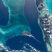
PI-RADS v2 Compliant Automated Segmentation of Prostate Zones Using co-training Motivated Multi-task Dual-Path CNN
The detailed images produced by Magnetic Resonance Imaging (MRI) provide life-critical information for the diagnosis and treatment of prostate cancer. To provide standardized acquisition, interpretation and usage of the complex MRI images, the PI-RADS v2 guideline was proposed. An automated segmentation following the guideline facilitates consistent and precise lesion detection, staging and treatment. The guideline recommends a division of the prostate into four zones, PZ (peripheral zone), TZ (transition zone), DPU (distal prostatic urethra) and AFS (anterior fibromuscular stroma). Not every zone shares a boundary with the others and is present in every slice. Further, the representations captured by a single model might not suffice for all zones. This motivated us to design a dual-branch convolutional neural network (CNN), where each branch captures the representations of the connected zones separately. Further, the representations from different branches act complementary to each other at the second stage of training, where they are fine-tuned through an unsupervised loss. The loss penalises the difference in predictions from the two branches for the same class. We also incorporate multi-task learning in our framework to further improve the segmentation accuracy. The proposed approach improves the segmentation accuracy of the baseline (mean absolute symmetric distance) by 7.56%, 11.00%, 58.43% and 19.67% for PZ, TZ, DPU and AFS zones respectively.
Generating Physically-Consistent Satellite Imagery for Climate Visualizations
Deep generative vision models are now able to synthesize realistic-looking satellite imagery. But, the possibility of hallucinations prevents their adoption for risk-sensitive applications, such as generating materials for communicating climate change. To demonstrate this issue, we train a generative adversarial network (pix2pixHD) to create synthetic satellite imagery of future flooding and reforestation events. We find that a pure deep learning-based model can generate photorealistic flood visualizations but hallucinates floods at locations that were not susceptible to flooding. To address this issue, we propose to condition and evaluate generative vision models on segmentation maps of physics-based flood models. We show that our physics-conditioned model outperforms the pure deep learning-based model and a handcrafted baseline. We evaluate the generalization capability of our method to different remote sensing data and different climate-related events (reforestation). We publish our code and dataset which includes the data for a third case study of melting Arctic sea ice and >30,000 labeled HD image triplets -- or the equivalent of 5.5 million images at 128x128 pixels -- for segmentation guided image-to-image translation in Earth observation. Code and data is available at https://github.com/blutjens/eie-earth-public.

SegVol: Universal and Interactive Volumetric Medical Image Segmentation
Precise image segmentation provides clinical study with meaningful and well-structured information. Despite the remarkable progress achieved in medical image segmentation, there is still an absence of foundation segmentation model that can segment a wide range of anatomical categories with easy user interaction. In this paper, we propose a universal and interactive volumetric medical image segmentation model, named SegVol. By training on 90k unlabeled Computed Tomography (CT) volumes and 6k labeled CTs, this foundation model supports the segmentation of over 200 anatomical categories using semantic and spatial prompts. Extensive experiments verify that SegVol outperforms the state of the art by a large margin on multiple segmentation benchmarks. Notably, on three challenging lesion datasets, our method achieves around 20% higher Dice score than nnU-Net. The model and data are publicly available at: https://github.com/BAAI-DCAI/SegVol.

SAModified: A Foundation Model-Based Zero-Shot Approach for Refining Noisy Land-Use Land-Cover Maps
Land-use and land cover (LULC) analysis is critical in remote sensing, with wide-ranging applications across diverse fields such as agriculture, utilities, and urban planning. However, automating LULC map generation using machine learning is rendered challenging due to noisy labels. Typically, the ground truths (e.g. ESRI LULC, MapBioMass) have noisy labels that hamper the model's ability to learn to accurately classify the pixels. Further, these erroneous labels can significantly distort the performance metrics of a model, leading to misleading evaluations. Traditionally, the ambiguous labels are rectified using unsupervised algorithms. These algorithms struggle not only with scalability but also with generalization across different geographies. To overcome these challenges, we propose a zero-shot approach using the foundation model, Segment Anything Model (SAM), to automatically delineate different land parcels/regions and leverage them to relabel the unsure pixels by using the local label statistics within each detected region. We achieve a significant reduction in label noise and an improvement in the performance of the downstream segmentation model by approx 5% when trained with denoised labels.
Point2Mask: Point-supervised Panoptic Segmentation via Optimal Transport
Weakly-supervised image segmentation has recently attracted increasing research attentions, aiming to avoid the expensive pixel-wise labeling. In this paper, we present an effective method, namely Point2Mask, to achieve high-quality panoptic prediction using only a single random point annotation per target for training. Specifically, we formulate the panoptic pseudo-mask generation as an Optimal Transport (OT) problem, where each ground-truth (gt) point label and pixel sample are defined as the label supplier and consumer, respectively. The transportation cost is calculated by the introduced task-oriented maps, which focus on the category-wise and instance-wise differences among the various thing and stuff targets. Furthermore, a centroid-based scheme is proposed to set the accurate unit number for each gt point supplier. Hence, the pseudo-mask generation is converted into finding the optimal transport plan at a globally minimal transportation cost, which can be solved via the Sinkhorn-Knopp Iteration. Experimental results on Pascal VOC and COCO demonstrate the promising performance of our proposed Point2Mask approach to point-supervised panoptic segmentation. Source code is available at: https://github.com/LiWentomng/Point2Mask.


Segmentation in large-scale cellular electron microscopy with deep learning: A literature survey
Automated and semi-automated techniques in biomedical electron microscopy (EM) enable the acquisition of large datasets at a high rate. Segmentation methods are therefore essential to analyze and interpret these large volumes of data, which can no longer completely be labeled manually. In recent years, deep learning algorithms achieved impressive results in both pixel-level labeling (semantic segmentation) and the labeling of separate instances of the same class (instance segmentation). In this review, we examine how these algorithms were adapted to the task of segmenting cellular and sub-cellular structures in EM images. The special challenges posed by such images and the network architectures that overcame some of them are described. Moreover, a thorough overview is also provided on the notable datasets that contributed to the proliferation of deep learning in EM. Finally, an outlook of current trends and future prospects of EM segmentation is given, especially in the area of label-free learning.
The Devil is in the Points: Weakly Semi-Supervised Instance Segmentation via Point-Guided Mask Representation
In this paper, we introduce a novel learning scheme named weakly semi-supervised instance segmentation (WSSIS) with point labels for budget-efficient and high-performance instance segmentation. Namely, we consider a dataset setting consisting of a few fully-labeled images and a lot of point-labeled images. Motivated by the main challenge of semi-supervised approaches mainly derives from the trade-off between false-negative and false-positive instance proposals, we propose a method for WSSIS that can effectively leverage the budget-friendly point labels as a powerful weak supervision source to resolve the challenge. Furthermore, to deal with the hard case where the amount of fully-labeled data is extremely limited, we propose a MaskRefineNet that refines noise in rough masks. We conduct extensive experiments on COCO and BDD100K datasets, and the proposed method achieves promising results comparable to those of the fully-supervised model, even with 50% of the fully labeled COCO data (38.8% vs. 39.7%). Moreover, when using as little as 5% of fully labeled COCO data, our method shows significantly superior performance over the state-of-the-art semi-supervised learning method (33.7% vs. 24.9%). The code is available at https://github.com/clovaai/PointWSSIS.

PathVG: A New Benchmark and Dataset for Pathology Visual Grounding
With the rapid development of computational pathology, many AI-assisted diagnostic tasks have emerged. Cellular nuclei segmentation can segment various types of cells for downstream analysis, but it relies on predefined categories and lacks flexibility. Moreover, pathology visual question answering can perform image-level understanding but lacks region-level detection capability. To address this, we propose a new benchmark called Pathology Visual Grounding (PathVG), which aims to detect regions based on expressions with different attributes. To evaluate PathVG, we create a new dataset named RefPath which contains 27,610 images with 33,500 language-grounded boxes. Compared to visual grounding in other domains, PathVG presents pathological images at multi-scale and contains expressions with pathological knowledge. In the experimental study, we found that the biggest challenge was the implicit information underlying the pathological expressions. Based on this, we proposed Pathology Knowledge-enhanced Network (PKNet) as the baseline model for PathVG. PKNet leverages the knowledge-enhancement capabilities of Large Language Models (LLMs) to convert pathological terms with implicit information into explicit visual features, and fuses knowledge features with expression features through the designed Knowledge Fusion Module (KFM). The proposed method achieves state-of-the-art performance on the PathVG benchmark.
Tiny and Efficient Model for the Edge Detection Generalization
Most high-level computer vision tasks rely on low-level image operations as their initial processes. Operations such as edge detection, image enhancement, and super-resolution, provide the foundations for higher level image analysis. In this work we address the edge detection considering three main objectives: simplicity, efficiency, and generalization since current state-of-the-art (SOTA) edge detection models are increased in complexity for better accuracy. To achieve this, we present Tiny and Efficient Edge Detector (TEED), a light convolutional neural network with only 58K parameters, less than 0.2% of the state-of-the-art models. Training on the BIPED dataset takes less than 30 minutes, with each epoch requiring less than 5 minutes. Our proposed model is easy to train and it quickly converges within very first few epochs, while the predicted edge-maps are crisp and of high quality. Additionally, we propose a new dataset to test the generalization of edge detection, which comprises samples from popular images used in edge detection and image segmentation. The source code is available in https://github.com/xavysp/TEED.
3D Medical Image Segmentation based on multi-scale MPU-Net
The high cure rate of cancer is inextricably linked to physicians' accuracy in diagnosis and treatment, therefore a model that can accomplish high-precision tumor segmentation has become a necessity in many applications of the medical industry. It can effectively lower the rate of misdiagnosis while considerably lessening the burden on clinicians. However, fully automated target organ segmentation is problematic due to the irregular stereo structure of 3D volume organs. As a basic model for this class of real applications, U-Net excels. It can learn certain global and local features, but still lacks the capacity to grasp spatial long-range relationships and contextual information at multiple scales. This paper proposes a tumor segmentation model MPU-Net for patient volume CT images, which is inspired by Transformer with a global attention mechanism. By combining image serialization with the Position Attention Module, the model attempts to comprehend deeper contextual dependencies and accomplish precise positioning. Each layer of the decoder is also equipped with a multi-scale module and a cross-attention mechanism. The capability of feature extraction and integration at different levels has been enhanced, and the hybrid loss function developed in this study can better exploit high-resolution characteristic information. Moreover, the suggested architecture is tested and evaluated on the Liver Tumor Segmentation Challenge 2017 (LiTS 2017) dataset. Compared with the benchmark model U-Net, MPU-Net shows excellent segmentation results. The dice, accuracy, precision, specificity, IOU, and MCC metrics for the best model segmentation results are 92.17%, 99.08%, 91.91%, 99.52%, 85.91%, and 91.74%, respectively. Outstanding indicators in various aspects illustrate the exceptional performance of this framework in automatic medical image segmentation.
The magnitude vector of images
The magnitude of a finite metric space has recently emerged as a novel invariant quantity, allowing to measure the effective size of a metric space. Despite encouraging first results demonstrating the descriptive abilities of the magnitude, such as being able to detect the boundary of a metric space, the potential use cases of magnitude remain under-explored. In this work, we investigate the properties of the magnitude on images, an important data modality in many machine learning applications. By endowing each individual images with its own metric space, we are able to define the concept of magnitude on images and analyse the individual contribution of each pixel with the magnitude vector. In particular, we theoretically show that the previously known properties of boundary detection translate to edge detection abilities in images. Furthermore, we demonstrate practical use cases of magnitude for machine learning applications and propose a novel magnitude model that consists of a computationally efficient magnitude computation and a learnable metric. By doing so, we address the computational hurdle that used to make magnitude impractical for many applications and open the way for the adoption of magnitude in machine learning research.
Learning to Synthesize a 4D RGBD Light Field from a Single Image
We present a machine learning algorithm that takes as input a 2D RGB image and synthesizes a 4D RGBD light field (color and depth of the scene in each ray direction). For training, we introduce the largest public light field dataset, consisting of over 3300 plenoptic camera light fields of scenes containing flowers and plants. Our synthesis pipeline consists of a convolutional neural network (CNN) that estimates scene geometry, a stage that renders a Lambertian light field using that geometry, and a second CNN that predicts occluded rays and non-Lambertian effects. Our algorithm builds on recent view synthesis methods, but is unique in predicting RGBD for each light field ray and improving unsupervised single image depth estimation by enforcing consistency of ray depths that should intersect the same scene point. Please see our supplementary video at https://youtu.be/yLCvWoQLnms

Synthetic Boost: Leveraging Synthetic Data for Enhanced Vision-Language Segmentation in Echocardiography
Accurate segmentation is essential for echocardiography-based assessment of cardiovascular diseases (CVDs). However, the variability among sonographers and the inherent challenges of ultrasound images hinder precise segmentation. By leveraging the joint representation of image and text modalities, Vision-Language Segmentation Models (VLSMs) can incorporate rich contextual information, potentially aiding in accurate and explainable segmentation. However, the lack of readily available data in echocardiography hampers the training of VLSMs. In this study, we explore using synthetic datasets from Semantic Diffusion Models (SDMs) to enhance VLSMs for echocardiography segmentation. We evaluate results for two popular VLSMs (CLIPSeg and CRIS) using seven different kinds of language prompts derived from several attributes, automatically extracted from echocardiography images, segmentation masks, and their metadata. Our results show improved metrics and faster convergence when pretraining VLSMs on SDM-generated synthetic images before finetuning on real images. The code, configs, and prompts are available at https://github.com/naamiinepal/synthetic-boost.

INT: Instance-Specific Negative Mining for Task-Generic Promptable Segmentation
Task-generic promptable image segmentation aims to achieve segmentation of diverse samples under a single task description by utilizing only one task-generic prompt. Current methods leverage the generalization capabilities of Vision-Language Models (VLMs) to infer instance-specific prompts from these task-generic prompts in order to guide the segmentation process. However, when VLMs struggle to generalise to some image instances, predicting instance-specific prompts becomes poor. To solve this problem, we introduce Instance-specific Negative Mining for Task-Generic Promptable Segmentation (INT). The key idea of INT is to adaptively reduce the influence of irrelevant (negative) prior knowledge whilst to increase the use the most plausible prior knowledge, selected by negative mining with higher contrast, in order to optimise instance-specific prompts generation. Specifically, INT consists of two components: (1) instance-specific prompt generation, which progressively fliters out incorrect information in prompt generation; (2) semantic mask generation, which ensures each image instance segmentation matches correctly the semantics of the instance-specific prompts. INT is validated on six datasets, including camouflaged objects and medical images, demonstrating its effectiveness, robustness and scalability.


U-DIADS-Bib: a full and few-shot pixel-precise dataset for document layout analysis of ancient manuscripts
Document Layout Analysis, which is the task of identifying different semantic regions inside of a document page, is a subject of great interest for both computer scientists and humanities scholars as it represents a fundamental step towards further analysis tasks for the former and a powerful tool to improve and facilitate the study of the documents for the latter. However, many of the works currently present in the literature, especially when it comes to the available datasets, fail to meet the needs of both worlds and, in particular, tend to lean towards the needs and common practices of the computer science side, leading to resources that are not representative of the humanities real needs. For this reason, the present paper introduces U-DIADS-Bib, a novel, pixel-precise, non-overlapping and noiseless document layout analysis dataset developed in close collaboration between specialists in the fields of computer vision and humanities. Furthermore, we propose a novel, computer-aided, segmentation pipeline in order to alleviate the burden represented by the time-consuming process of manual annotation, necessary for the generation of the ground truth segmentation maps. Finally, we present a standardized few-shot version of the dataset (U-DIADS-BibFS), with the aim of encouraging the development of models and solutions able to address this task with as few samples as possible, which would allow for more effective use in a real-world scenario, where collecting a large number of segmentations is not always feasible.

A smartphone application to measure the quality of pest control spraying machines via image analysis
The need for higher agricultural productivity has demanded the intensive use of pesticides. However, their correct use depends on assessment methods that can accurately predict how well the pesticides' spraying covered the intended crop region. Some methods have been proposed in the literature, but their high cost and low portability harm their widespread use. This paper proposes and experimentally evaluates a new methodology based on the use of a smartphone-based mobile application, named DropLeaf. Experiments performed using DropLeaf showed that, in addition to its versatility, it can predict with high accuracy the pesticide spraying. DropLeaf is a five-fold image-processing methodology based on: (i) color space conversion, (ii) threshold noise removal, (iii) convolutional operations of dilation and erosion, (iv) detection of contour markers in the water-sensitive card, and, (v) identification of droplets via the marker-controlled watershed transformation. The authors performed successful experiments over two case studies, the first using a set of synthetic cards and the second using a real-world crop. The proposed tool can be broadly used by farmers equipped with conventional mobile phones, improving the use of pesticides with health, environmental and financial benefits.
Tracking Anything with Decoupled Video Segmentation
Training data for video segmentation are expensive to annotate. This impedes extensions of end-to-end algorithms to new video segmentation tasks, especially in large-vocabulary settings. To 'track anything' without training on video data for every individual task, we develop a decoupled video segmentation approach (DEVA), composed of task-specific image-level segmentation and class/task-agnostic bi-directional temporal propagation. Due to this design, we only need an image-level model for the target task (which is cheaper to train) and a universal temporal propagation model which is trained once and generalizes across tasks. To effectively combine these two modules, we use bi-directional propagation for (semi-)online fusion of segmentation hypotheses from different frames to generate a coherent segmentation. We show that this decoupled formulation compares favorably to end-to-end approaches in several data-scarce tasks including large-vocabulary video panoptic segmentation, open-world video segmentation, referring video segmentation, and unsupervised video object segmentation. Code is available at: https://hkchengrex.github.io/Tracking-Anything-with-DEVA


MLP Can Be A Good Transformer Learner
Self-attention mechanism is the key of the Transformer but often criticized for its computation demands. Previous token pruning works motivate their methods from the view of computation redundancy but still need to load the full network and require same memory costs. This paper introduces a novel strategy that simplifies vision transformers and reduces computational load through the selective removal of non-essential attention layers, guided by entropy considerations. We identify that regarding the attention layer in bottom blocks, their subsequent MLP layers, i.e. two feed-forward layers, can elicit the same entropy quantity. Meanwhile, the accompanied MLPs are under-exploited since they exhibit smaller feature entropy compared to those MLPs in the top blocks. Therefore, we propose to integrate the uninformative attention layers into their subsequent counterparts by degenerating them into identical mapping, yielding only MLP in certain transformer blocks. Experimental results on ImageNet-1k show that the proposed method can remove 40% attention layer of DeiT-B, improving throughput and memory bound without performance compromise. Code is available at https://github.com/sihaoevery/lambda_vit.
DIAMANT: Dual Image-Attention Map Encoders For Medical Image Segmentation
Although purely transformer-based architectures showed promising performance in many computer vision tasks, many hybrid models consisting of CNN and transformer blocks are introduced to fit more specialized tasks. Nevertheless, despite the performance gain of both pure and hybrid transformer-based architectures compared to CNNs in medical imaging segmentation, their high training cost and complexity make it challenging to use them in real scenarios. In this work, we propose simple architectures based on purely convolutional layers, and show that by just taking advantage of the attention map visualizations obtained from a self-supervised pretrained vision transformer network (e.g., DINO) one can outperform complex transformer-based networks with much less computation costs. The proposed architecture is composed of two encoder branches with the original image as input in one branch and the attention map visualizations of the same image from multiple self-attention heads from a pre-trained DINO model (as multiple channels) in the other branch. The results of our experiments on two publicly available medical imaging datasets show that the proposed pipeline outperforms U-Net and the state-of-the-art medical image segmentation models.
POBEVM: Real-time Video Matting via Progressively Optimize the Target Body and Edge
Deep convolutional neural networks (CNNs) based approaches have achieved great performance in video matting. Many of these methods can produce accurate alpha estimation for the target body but typically yield fuzzy or incorrect target edges. This is usually caused by the following reasons: 1) The current methods always treat the target body and edge indiscriminately; 2) Target body dominates the whole target with only a tiny proportion target edge. For the first problem, we propose a CNN-based module that separately optimizes the matting target body and edge (SOBE). And on this basis, we introduce a real-time, trimap-free video matting method via progressively optimizing the matting target body and edge (POBEVM) that is much lighter than previous approaches and achieves significant improvements in the predicted target edge. For the second problem, we propose an Edge-L1-Loss (ELL) function that enforces our network on the matting target edge. Experiments demonstrate our method outperforms prior trimap-free matting methods on both Distinctions-646 (D646) and VideoMatte240K(VM) dataset, especially in edge optimization.
Tumor Detection, Segmentation and Classification Challenge on Automated 3D Breast Ultrasound: The TDSC-ABUS Challenge
Breast cancer is one of the most common causes of death among women worldwide. Early detection helps in reducing the number of deaths. Automated 3D Breast Ultrasound (ABUS) is a newer approach for breast screening, which has many advantages over handheld mammography such as safety, speed, and higher detection rate of breast cancer. Tumor detection, segmentation, and classification are key components in the analysis of medical images, especially challenging in the context of 3D ABUS due to the significant variability in tumor size and shape, unclear tumor boundaries, and a low signal-to-noise ratio. The lack of publicly accessible, well-labeled ABUS datasets further hinders the advancement of systems for breast tumor analysis. Addressing this gap, we have organized the inaugural Tumor Detection, Segmentation, and Classification Challenge on Automated 3D Breast Ultrasound 2023 (TDSC-ABUS2023). This initiative aims to spearhead research in this field and create a definitive benchmark for tasks associated with 3D ABUS image analysis. In this paper, we summarize the top-performing algorithms from the challenge and provide critical analysis for ABUS image examination. We offer the TDSC-ABUS challenge as an open-access platform at https://tdsc-abus2023.grand-challenge.org/ to benchmark and inspire future developments in algorithmic research.
Effortless Efficiency: Low-Cost Pruning of Diffusion Models
Diffusion models have achieved impressive advancements in various vision tasks. However, these gains often rely on increasing model size, which escalates computational complexity and memory demands, complicating deployment, raising inference costs, and causing environmental impact. While some studies have explored pruning techniques to improve the memory efficiency of diffusion models, most existing methods require extensive retraining to retain the model performance. Retraining a modern large diffusion model is extremely costly and resource-intensive, which limits the practicality of these methods. In this work, we achieve low-cost diffusion pruning without retraining by proposing a model-agnostic structural pruning framework for diffusion models that learns a differentiable mask to sparsify the model. To ensure effective pruning that preserves the quality of the final denoised latent, we design a novel end-to-end pruning objective that spans the entire diffusion process. As end-to-end pruning is memory-intensive, we further propose time step gradient checkpointing, a technique that significantly reduces memory usage during optimization, enabling end-to-end pruning within a limited memory budget. Results on state-of-the-art U-Net diffusion models SDXL and diffusion transformers (FLUX) demonstrate that our method can effectively prune up to 20% parameters with minimal perceptible performance degradation, and notably, without the need for model retraining. We also showcase that our method can still prune on top of time step distilled diffusion models.
PairingNet: A Learning-based Pair-searching and -matching Network for Image Fragments
In this paper, we propose a learning-based image fragment pair-searching and -matching approach to solve the challenging restoration problem. Existing works use rule-based methods to match similar contour shapes or textures, which are always difficult to tune hyperparameters for extensive data and computationally time-consuming. Therefore, we propose a neural network that can effectively utilize neighbor textures with contour shape information to fundamentally improve performance. First, we employ a graph-based network to extract the local contour and texture features of fragments. Then, for the pair-searching task, we adopt a linear transformer-based module to integrate these local features and use contrastive loss to encode the global features of each fragment. For the pair-matching task, we design a weighted fusion module to dynamically fuse extracted local contour and texture features, and formulate a similarity matrix for each pair of fragments to calculate the matching score and infer the adjacent segment of contours. To faithfully evaluate our proposed network, we created a new image fragment dataset through an algorithm we designed that tears complete images into irregular fragments. The experimental results show that our proposed network achieves excellent pair-searching accuracy, reduces matching errors, and significantly reduces computational time. Details, sourcecode, and data are available in our supplementary material.
View-Consistent Hierarchical 3D Segmentation Using Ultrametric Feature Fields
Large-scale vision foundation models such as Segment Anything (SAM) demonstrate impressive performance in zero-shot image segmentation at multiple levels of granularity. However, these zero-shot predictions are rarely 3D-consistent. As the camera viewpoint changes in a scene, so do the segmentation predictions, as well as the characterizations of "coarse" or "fine" granularity. In this work, we address the challenging task of lifting multi-granular and view-inconsistent image segmentations into a hierarchical and 3D-consistent representation. We learn a novel feature field within a Neural Radiance Field (NeRF) representing a 3D scene, whose segmentation structure can be revealed at different scales by simply using different thresholds on feature distance. Our key idea is to learn an ultrametric feature space, which unlike a Euclidean space, exhibits transitivity in distance-based grouping, naturally leading to a hierarchical clustering. Put together, our method takes view-inconsistent multi-granularity 2D segmentations as input and produces a hierarchy of 3D-consistent segmentations as output. We evaluate our method and several baselines on synthetic datasets with multi-view images and multi-granular segmentation, showcasing improved accuracy and viewpoint-consistency. We additionally provide qualitative examples of our model's 3D hierarchical segmentations in real world scenes. The code and dataset are available at https://github.com/hardyho/ultrametric_feature_fields

Mask2Former for Video Instance Segmentation
We find Mask2Former also achieves state-of-the-art performance on video instance segmentation without modifying the architecture, the loss or even the training pipeline. In this report, we show universal image segmentation architectures trivially generalize to video segmentation by directly predicting 3D segmentation volumes. Specifically, Mask2Former sets a new state-of-the-art of 60.4 AP on YouTubeVIS-2019 and 52.6 AP on YouTubeVIS-2021. We believe Mask2Former is also capable of handling video semantic and panoptic segmentation, given its versatility in image segmentation. We hope this will make state-of-the-art video segmentation research more accessible and bring more attention to designing universal image and video segmentation architectures.